 Movie
Movie Controller

[English] 日本語
 Yorodumi
- PDB-6xrv: X-ray structure of the monoclinic crystal form at 1.43 A resoluti... -
+ Open data

- Basic information
| Entry | Database: PDB / ID: 6xrv | ||||||
|---|---|---|---|---|---|---|---|
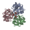
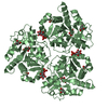
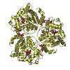
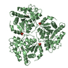
| Title | X-ray structure of the monoclinic crystal form at 1.43 A resolution of lipase from Thermomyces (Humicola) lanuginosa at 173 K | ||||||
 Components Components | Lipase | ||||||
 Keywords Keywords | LIPID BINDING PROTEIN / substrate complex / diacylglycerol / covalent intermediate / interfacial activation | ||||||
| Function / homology |  Function and homology information Function and homology informationtriacylglycerol lipase / triacylglycerol lipase activity / lipid catabolic process Similarity search - Function | ||||||
| Biological species |   Thermomyces lanuginosus (fungus) Thermomyces lanuginosus (fungus) | ||||||
| Method |  X-RAY DIFFRACTION / SYNCHROTRON / MOLECULAR REPLACEMENT / Resolution: 1.43 Å X-RAY DIFFRACTION / SYNCHROTRON / MOLECULAR REPLACEMENT / Resolution: 1.43 Å | ||||||
 Authors Authors | McPherson, A. | ||||||
 Citation Citation | Journal: Current Enzyme Inhibition / Year: 2020 Title: The crystal Structures of Thermomyces (Humicola) lanuginosa lipase in complex with enzymatic reactants Authors: McPherson, A. / Larson, S.B. / Kalasky, A. | ||||||
| History |
|
- Structure visualization
| Structure viewer | Molecule: MolmilJmol/JSmol |
|---|
- Downloads & links
-Download
| PDBx/mmCIF format | 6xrv.cif.gz | 980.5 KB | Display | PDBx/mmCIF format |
|---|---|---|---|---|
| PDB format | pdb6xrv.ent.gz | 833 KB | Display | PDB format |
| PDBx/mmJSON format | 6xrv.json.gz | Tree view | PDBx/mmJSON format | |
| Others |  Other downloads Other downloads |
-Validation report
| Arichive directory | https://data.pdbj.org/pub/pdb/validation_reports/xr/6xrvftp://data.pdbj.org/pub/pdb/validation_reports/xr/6xrv | HTTPS FTP |
|---|
-Related structure data
| Related structure data |  6or3C  6xokC  6xs3C  1tibS S: Starting model for refinement C: citing same article ( |
|---|---|
| Similar structure data |


-Links
 PDBj
PDBj- Assembly
| Deposited unit | 
| ||||||||
|---|---|---|---|---|---|---|---|---|---|
| 1 | 
| ||||||||
| 2 | 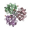
| ||||||||
| Unit cell |
|
-Components
-Protein / Sugars , 2 types, 12 molecules ABCEDF
| #1: Protein | Mass: 31836.459 Da / Num. of mol.: 6 Source method: isolated from a genetically manipulated source Source: (gene. exp.) Thermomyces lanuginosus (fungus) / Gene: LIP / Production host: #3: Sugar | ChemComp-NAG /  Type: D-saccharide, beta linking / Mass: 221.208 Da / Num. of mol.: 6 Type: D-saccharide, beta linking / Mass: 221.208 Da / Num. of mol.: 6Source method: isolated from a genetically manipulated source Formula: C8H15NO6 |
|---|
-Non-polymers , 5 types, 1645 molecules 


| #2: Chemical | ChemComp-OCA /  Mass: 144.211 Da / Num. of mol.: 6 / Source method: obtained synthetically / Formula: C8H16O2 Mass: 144.211 Da / Num. of mol.: 6 / Source method: obtained synthetically / Formula: C8H16O2#4: Chemical | ChemComp-LTV /  Mass: 723.204 Da / Num. of mol.: 6 / Source method: obtained synthetically / Formula: C46H90O5 / Feature type: SUBJECT OF INVESTIGATION Mass: 723.204 Da / Num. of mol.: 6 / Source method: obtained synthetically / Formula: C46H90O5 / Feature type: SUBJECT OF INVESTIGATION#5: Chemical | ChemComp-PO4 /  Mass: 94.971 Da / Num. of mol.: 9 / Source method: obtained synthetically / Formula: PO4 Mass: 94.971 Da / Num. of mol.: 9 / Source method: obtained synthetically / Formula: PO4#6: Chemical | ChemComp-CA /  Mass: 40.078 Da / Num. of mol.: 6 / Source method: obtained synthetically / Formula: Ca Mass: 40.078 Da / Num. of mol.: 6 / Source method: obtained synthetically / Formula: Ca#7: Water | ChemComp-HOH / | Mass: 18.015 Da / Num. of mol.: 1618 / Source method: isolated from a natural source / Formula: H2O |
|---|
-Details
| Has ligand of interest | Y |
|---|---|
| Has protein modification | Y |
-Experimental details
-Experiment
| Experiment | Method: X-RAY DIFFRACTION / Number of used crystals: 1 |
|---|
- Sample preparation
| Crystal | Density Matthews: 2.23 Å3/Da / Density % sol: 44.8 % / Description: cube like blocks |
|---|---|
| Crystal grow | Temperature: 295 K / Method: vapor diffusion, sitting drop / pH: 6.5 Details: Crystals grown by sitting drop with 0.6 ul reservoirs of 20% PEG 3350 in 0.1 M MES buffer at pH 6.5. Drops of 8 ul composed of equal amounts of the protein at 30.0 mg/ml in water with the ...Details: Crystals grown by sitting drop with 0.6 ul reservoirs of 20% PEG 3350 in 0.1 M MES buffer at pH 6.5. Drops of 8 ul composed of equal amounts of the protein at 30.0 mg/ml in water with the reservoir solution. Room temperature throughout, crystals grew in about a month PH range: 6.0 - 7.0 |
-Data collection
| Diffraction | Mean temperature: 173 K / Serial crystal experiment: N |
|---|---|
| Diffraction source | Source: SYNCHROTRON / Site: ALS  / Beamline: 8.3.1 / Wavelength: 1 Å / Beamline: 8.3.1 / Wavelength: 1 Å |
| Detector | Type: DECTRIS PILATUS 300K / Detector: PIXEL / Date: Feb 22, 2019 |
| Radiation | Protocol: SINGLE WAVELENGTH / Monochromatic (M) / Laue (L): M / Scattering type: x-ray |
| Radiation wavelength | Wavelength: 1 Å / Relative weight: 1 |
| Reflection | Resolution: 1.43→77 Å / Num. obs: 307462 / % possible obs: 99.7 % / Redundancy: 19.2 % / Biso Wilson estimate: 22 Å2 / CC1/2: 0.998 / Rmerge(I) obs: 0.158 / Rpim(I) all: 0.036 / Rrim(I) all: 0.163 / Rsym value: 0.156 / Net I/σ(I): 9.1 |
| Reflection shell | Resolution: 1.43→1.45 Å / Redundancy: 13.3 % / Rmerge(I) obs: 5.69 / Mean I/σ(I) obs: 0.4 / Num. unique obs: 15076 / CC1/2: 0.26 / Rpim(I) all: 1.66 / Rrim(I) all: 6.1 / % possible all: 98.8 |
- Processing
| Software |
| |||||||||||||||||||||||||||||||||||||||||||||||||||||||||||||||||||||||||||||||||||||||||||||||||||||||||||||||||||||||||||||||||||||||||||||||||||||||||||||||||||||||||||||||||||||||||||||||||||||||||||||||||||||||||
|---|---|---|---|---|---|---|---|---|---|---|---|---|---|---|---|---|---|---|---|---|---|---|---|---|---|---|---|---|---|---|---|---|---|---|---|---|---|---|---|---|---|---|---|---|---|---|---|---|---|---|---|---|---|---|---|---|---|---|---|---|---|---|---|---|---|---|---|---|---|---|---|---|---|---|---|---|---|---|---|---|---|---|---|---|---|---|---|---|---|---|---|---|---|---|---|---|---|---|---|---|---|---|---|---|---|---|---|---|---|---|---|---|---|---|---|---|---|---|---|---|---|---|---|---|---|---|---|---|---|---|---|---|---|---|---|---|---|---|---|---|---|---|---|---|---|---|---|---|---|---|---|---|---|---|---|---|---|---|---|---|---|---|---|---|---|---|---|---|---|---|---|---|---|---|---|---|---|---|---|---|---|---|---|---|---|---|---|---|---|---|---|---|---|---|---|---|---|---|---|---|---|---|---|---|---|---|---|---|---|---|---|---|---|---|---|---|---|---|
| Refinement | Method to determine structure: MOLECULAR REPLACEMENT Starting model: 1TIB Resolution: 1.43→77 Å / SU ML: 0.19 / Cross valid method: THROUGHOUT / σ(F): 1.33 / Phase error: 19.66 / Stereochemistry target values: ML Details: Refined to convergence in REFMAC then subjected to further rebuilding in COOT and an additional 15 runs in REFINE from PHENIX. Anisotropic B values, no TLS.
| |||||||||||||||||||||||||||||||||||||||||||||||||||||||||||||||||||||||||||||||||||||||||||||||||||||||||||||||||||||||||||||||||||||||||||||||||||||||||||||||||||||||||||||||||||||||||||||||||||||||||||||||||||||||||
| Solvent computation | Shrinkage radii: 0.9 Å / VDW probe radii: 1.11 Å / Solvent model: FLAT BULK SOLVENT MODEL | |||||||||||||||||||||||||||||||||||||||||||||||||||||||||||||||||||||||||||||||||||||||||||||||||||||||||||||||||||||||||||||||||||||||||||||||||||||||||||||||||||||||||||||||||||||||||||||||||||||||||||||||||||||||||
| Displacement parameters | Biso max: 74.3 Å2 / Biso mean: 29.7343 Å2 / Biso min: 8.46 Å2 | |||||||||||||||||||||||||||||||||||||||||||||||||||||||||||||||||||||||||||||||||||||||||||||||||||||||||||||||||||||||||||||||||||||||||||||||||||||||||||||||||||||||||||||||||||||||||||||||||||||||||||||||||||||||||
| Refinement step | Cycle: final / Resolution: 1.43→77 Å
| |||||||||||||||||||||||||||||||||||||||||||||||||||||||||||||||||||||||||||||||||||||||||||||||||||||||||||||||||||||||||||||||||||||||||||||||||||||||||||||||||||||||||||||||||||||||||||||||||||||||||||||||||||||||||
| LS refinement shell | Refine-ID: X-RAY DIFFRACTION / Rfactor Rfree error: 0 / Total num. of bins used: 30
|
