 Movie
Movie Controller


+ Open data

- Basic information
| Entry | Database: PDB / ID: 6xhp | ||||||||||||
|---|---|---|---|---|---|---|---|---|---|---|---|---|---|
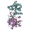
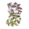
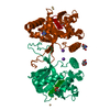
| Title | Crystal structure of S. aureus TarI (space group C121) | ||||||||||||
 Components Components | Ribitol-5-phosphate cytidylyltransferase 1 | ||||||||||||
 Keywords Keywords | TRANSFERASE / cytidylyltransferase | ||||||||||||
| Function / homology |  Function and homology information Function and homology informationD-ribitol-5-phosphate cytidylyltransferase / D-ribitol-5-phosphate cytidylyltransferase activity / poly(ribitol phosphate) teichoic acid biosynthetic process / 2-C-methyl-D-erythritol 4-phosphate cytidylyltransferase activity / isoprenoid biosynthetic process / cell wall organization Similarity search - Function | ||||||||||||
| Biological species |   Staphylococcus aureus (bacteria) Staphylococcus aureus (bacteria) | ||||||||||||
| Method |  X-RAY DIFFRACTION / SYNCHROTRON / MOLECULAR REPLACEMENT / Resolution: 1.9 Å X-RAY DIFFRACTION / SYNCHROTRON / MOLECULAR REPLACEMENT / Resolution: 1.9 Å | ||||||||||||
 Authors Authors | Li, F.K.K. / Strynadka, N.C.J. | ||||||||||||
| Funding support |  Canada, 3items Canada, 3items
| ||||||||||||
 Citation Citation | Journal: J.Struct.Biol. / Year: 2021 Title: Crystallographic analysis of TarI and TarJ, a cytidylyltransferase and reductase pair for CDP-ribitol synthesis in Staphylococcus aureus wall teichoic acid biogenesis. Authors: Li, F.K.K. / Gale, R.T. / Petrotchenko, E.V. / Borchers, C.H. / Brown, E.D. / Strynadka, N.C.J. | ||||||||||||
| History |
|
- Structure visualization
| Structure viewer | Molecule: MolmilJmol/JSmol |
|---|
- Downloads & links
-Download
| PDBx/mmCIF format | 6xhp.cif.gz | 195.8 KB | Display | PDBx/mmCIF format |
|---|---|---|---|---|
| PDB format | pdb6xhp.ent.gz | 154 KB | Display | PDB format |
| PDBx/mmJSON format | 6xhp.json.gz | Tree view | PDBx/mmJSON format | |
| Others |  Other downloads Other downloads |
-Validation report
| Arichive directory | https://data.pdbj.org/pub/pdb/validation_reports/xh/6xhpftp://data.pdbj.org/pub/pdb/validation_reports/xh/6xhp | HTTPS FTP |
|---|
-Related structure data
| Related structure data | 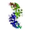 6xh9C 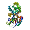 6xhkC 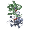 6xhqC 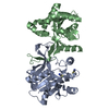 6xhrC 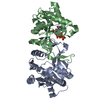 6xhsC 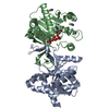 6xhtC 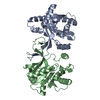 4jisS S: Starting model for refinement C: citing same article ( |
|---|---|
| Similar structure data |


-Links
 PDBj
PDBj

- Assembly
| Deposited unit | 
| ||||||||||||||||||||||||||||||||||||||||||||||||||||||||||||||||||||||||||||||||||||||||||
|---|---|---|---|---|---|---|---|---|---|---|---|---|---|---|---|---|---|---|---|---|---|---|---|---|---|---|---|---|---|---|---|---|---|---|---|---|---|---|---|---|---|---|---|---|---|---|---|---|---|---|---|---|---|---|---|---|---|---|---|---|---|---|---|---|---|---|---|---|---|---|---|---|---|---|---|---|---|---|---|---|---|---|---|---|---|---|---|---|---|---|---|
| 1 |
| ||||||||||||||||||||||||||||||||||||||||||||||||||||||||||||||||||||||||||||||||||||||||||
| Unit cell |
| ||||||||||||||||||||||||||||||||||||||||||||||||||||||||||||||||||||||||||||||||||||||||||
| Noncrystallographic symmetry (NCS) | NCS domain:
NCS domain segments: Ens-ID: 1
|