 Movie
Movie Controller

[English] 日本語
 Yorodumi
- PDB-4jis: Crystal structure of ribitol 5-phosphate cytidylyltransferase (Ta... -
+ Open data

- Basic information
| Entry | Database: PDB / ID: 4jis | ||||||
|---|---|---|---|---|---|---|---|
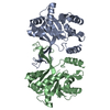
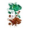
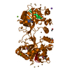
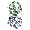
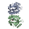
| Title | Crystal structure of ribitol 5-phosphate cytidylyltransferase (TarI) from Bacillus subtilis | ||||||
 Components Components | ribitol-5-phosphate cytidylyltransferase | ||||||
 Keywords Keywords | TRANSFERASE / ribitol 5-phosphate cytidylyltransferase | ||||||
| Function / homology |  Function and homology information Function and homology informationD-ribitol-5-phosphate cytidylyltransferase / D-ribitol-5-phosphate cytidylyltransferase activity / poly(ribitol phosphate) teichoic acid biosynthetic process / 2-C-methyl-D-erythritol 4-phosphate cytidylyltransferase activity / isoprenoid biosynthetic process / cell wall organization Similarity search - Function | ||||||
| Biological species |  | ||||||
| Method |  X-RAY DIFFRACTION / SYNCHROTRON / MOLECULAR REPLACEMENT / Resolution: 1.772 Å X-RAY DIFFRACTION / SYNCHROTRON / MOLECULAR REPLACEMENT / Resolution: 1.772 Å | ||||||
 Authors Authors | Yang, C.S. / Chen, S.C. / Chen, Y.R. / Kuan, S.M. / Liu, Y.H. / Chen, Y. | ||||||
 Citation Citation | Journal: To be Published Title: Crystal structure of ribitol 5-phosphate cytidylyltransferase (TarI) from Bacillus subtilis Authors: Yang, C.S. / Chen, S.C. / Chen, Y.R. / Kuan, S.M. / Liu, Y.H. / Chen, Y. | ||||||
| History |
|
- Structure visualization
| Structure viewer | Molecule: MolmilJmol/JSmol |
|---|
- Downloads & links
-Download
| PDBx/mmCIF format | 4jis.cif.gz | 107.4 KB | Display | PDBx/mmCIF format |
|---|---|---|---|---|
| PDB format | pdb4jis.ent.gz | 81.9 KB | Display | PDB format |
| PDBx/mmJSON format | 4jis.json.gz | Tree view | PDBx/mmJSON format | |
| Others |  Other downloads Other downloads |
-Validation report
| Arichive directory | https://data.pdbj.org/pub/pdb/validation_reports/ji/4jisftp://data.pdbj.org/pub/pdb/validation_reports/ji/4jis | HTTPS FTP |
|---|
-Related structure data
| Related structure data |  3f1cS S: Starting model for refinement |
|---|---|
| Similar structure data |


-Links
 PDBj
PDBj

- Assembly
| Deposited unit | 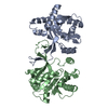
| ||||||||
|---|---|---|---|---|---|---|---|---|---|
| 1 |
| ||||||||
| Unit cell |
|
-Components
| #1: Protein | Mass: 28141.354 Da / Num. of mol.: 2 Source method: isolated from a genetically manipulated source Source: (gene. exp.) Strain: w23 / Gene: BSUW23_17565, ispD, tarI / Plasmid: pET28a(+) / Production host: References: UniProt: Q8RKI9, D-ribitol-5-phosphate cytidylyltransferase #2: Water | ChemComp-HOH / |  Mass: 18.015 Da / Num. of mol.: 325 / Source method: isolated from a natural source / Formula: H2O Mass: 18.015 Da / Num. of mol.: 325 / Source method: isolated from a natural source / Formula: H2O |
|---|
-Experimental details
-Experiment
| Experiment | Method: X-RAY DIFFRACTION / Number of used crystals: 1 |
|---|
- Sample preparation
| Crystal | Density Matthews: 2.37 Å3/Da / Density % sol: 48.03 % |
|---|---|
| Crystal grow | Temperature: 277 K / Method: vapor diffusion, sitting drop / pH: 6 Details: 20% PEG 6000, 0.1M MES-NaOH (pH 6.0), 0.2M CaCl2, VAPOR DIFFUSION, SITTING DROP, temperature 277K |
-Data collection
| Diffraction | Mean temperature: 173 K |
|---|---|
| Diffraction source | Source: SYNCHROTRON / Site: NSRRC  / Beamline: BL13C1 / Wavelength: 1 Å / Beamline: BL13C1 / Wavelength: 1 Å |
| Detector | Type: ADSC QUANTUM 315r / Detector: CCD / Date: Mar 30, 2012 |
| Radiation | Monochromator: Si 111 CHANNEL / Protocol: SINGLE WAVELENGTH / Monochromatic (M) / Laue (L): M / Scattering type: x-ray |
| Radiation wavelength | Wavelength: 1 Å / Relative weight: 1 |
| Reflection | Resolution: 1.772→30 Å / Num. all: 50947 / Num. obs: 49878 / % possible obs: 97.9 % / Observed criterion σ(F): 0 / Observed criterion σ(I): 0 |
| Reflection shell | Resolution: 1.78→1.84 Å / Redundancy: 4.5 % / Rmerge(I) obs: 0.458 / Num. unique all: 4894 / % possible all: 96.8 |
- Processing
| Software |
| ||||||||||||||||||||||||||||||||||||||||||||||||||||||||||||||||||||||||||||||||||||||||||||||||||||||||||||||||||
|---|---|---|---|---|---|---|---|---|---|---|---|---|---|---|---|---|---|---|---|---|---|---|---|---|---|---|---|---|---|---|---|---|---|---|---|---|---|---|---|---|---|---|---|---|---|---|---|---|---|---|---|---|---|---|---|---|---|---|---|---|---|---|---|---|---|---|---|---|---|---|---|---|---|---|---|---|---|---|---|---|---|---|---|---|---|---|---|---|---|---|---|---|---|---|---|---|---|---|---|---|---|---|---|---|---|---|---|---|---|---|---|---|---|---|---|
| Refinement | Method to determine structure: MOLECULAR REPLACEMENT Starting model: 3F1C Resolution: 1.772→24.71 Å / Occupancy max: 1 / Occupancy min: 1 / FOM work R set: 0.8156 / SU ML: 0.18 / σ(F): 1.37 / Phase error: 25.77 / Stereochemistry target values: ML
| ||||||||||||||||||||||||||||||||||||||||||||||||||||||||||||||||||||||||||||||||||||||||||||||||||||||||||||||||||
| Solvent computation | Shrinkage radii: 1 Å / VDW probe radii: 1.2 Å / Solvent model: FLAT BULK SOLVENT MODEL | ||||||||||||||||||||||||||||||||||||||||||||||||||||||||||||||||||||||||||||||||||||||||||||||||||||||||||||||||||
| Displacement parameters | Biso max: 77.25 Å2 / Biso mean: 38.96 Å2 / Biso min: 11.6 Å2 | ||||||||||||||||||||||||||||||||||||||||||||||||||||||||||||||||||||||||||||||||||||||||||||||||||||||||||||||||||
| Refinement step | Cycle: LAST / Resolution: 1.772→24.71 Å
| ||||||||||||||||||||||||||||||||||||||||||||||||||||||||||||||||||||||||||||||||||||||||||||||||||||||||||||||||||
| Refine LS restraints |
| ||||||||||||||||||||||||||||||||||||||||||||||||||||||||||||||||||||||||||||||||||||||||||||||||||||||||||||||||||
| LS refinement shell | Refine-ID: X-RAY DIFFRACTION / Total num. of bins used: 18
|
