| Entry | Database: PDB / ID: 4mkn
|
|---|
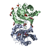
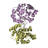
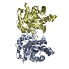
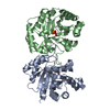
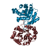
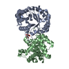
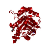
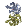
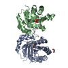
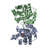
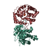
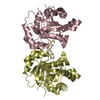
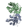
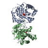
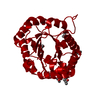
| Title | Crystal structure of chloroplastic triosephosphate isomerase from Chlamydomonas reinhardtii at 1.1 A of resolution |
|---|
 Components Components | Triosephosphate isomerase |
|---|
 Keywords Keywords | ISOMERASE / TIM barrel / chloroplast |
|---|
| Function / homology |  Function and homology information Function and homology information
triose-phosphate isomerase activity / glyceraldehyde-3-phosphate biosynthetic process / glycerol catabolic process / glycolytic process / gluconeogenesis / cytosolSimilarity search - Function Triosephosphate isomerase, bacterial/eukaryotic / Triosephosphate isomerase, active site / Triosephosphate isomerase active site. / Triosephosphate isomerase / Triosephosphate isomerase superfamily / Triosephosphate isomerase / Triosephosphate isomerase (TIM) family profile. / Aldolase class I / Aldolase-type TIM barrel / TIM Barrel ...Triosephosphate isomerase, bacterial/eukaryotic / Triosephosphate isomerase, active site / Triosephosphate isomerase active site. / Triosephosphate isomerase / Triosephosphate isomerase superfamily / Triosephosphate isomerase / Triosephosphate isomerase (TIM) family profile. / Aldolase class I / Aldolase-type TIM barrel / TIM Barrel / Alpha-Beta Barrel / Alpha BetaSimilarity search - Domain/homology |
|---|
| Biological species |   Chlamydomonas reinhardtii (plant) Chlamydomonas reinhardtii (plant) |
|---|
| Method |  X-RAY DIFFRACTION / SYNCHROTRON / MOLECULAR REPLACEMENT / Resolution: 1.1 Å X-RAY DIFFRACTION / SYNCHROTRON / MOLECULAR REPLACEMENT / Resolution: 1.1 Å |
|---|
 Authors Authors | Fermani, S. / Sciabolini, C. / Zaffagnini, M. / Lemaire, S.D. |
|---|
 Citation Citation | Journal: Mol Plant / Year: 2014
Title: High-Resolution Crystal Structure and Redox Properties of Chloroplastic Triosephosphate Isomerase from Chlamydomonas reinhardtii.
Authors: Zaffagnini, M. / Michelet, L. / Sciabolini, C. / Di Giacinto, N. / Morisse, S. / Marchand, C.H. / Trost, P. / Fermani, S. / Lemaire, S.D. |
|---|
| History | | Deposition | Sep 5, 2013 | Deposition site: RCSB / Processing site: RCSB |
|---|
| Revision 1.0 | Jan 1, 2014 | Provider: repository / Type: Initial release |
|---|
| Revision 1.1 | Jan 15, 2014 | Group: Database references |
|---|
| Revision 1.2 | Sep 20, 2023 | Group: Data collection / Database references ...Data collection / Database references / Derived calculations / Refinement description
Category: chem_comp_atom / chem_comp_bond ...chem_comp_atom / chem_comp_bond / database_2 / pdbx_initial_refinement_model / struct_site
Item: _database_2.pdbx_DOI / _database_2.pdbx_database_accession ..._database_2.pdbx_DOI / _database_2.pdbx_database_accession / _struct_site.pdbx_auth_asym_id / _struct_site.pdbx_auth_comp_id / _struct_site.pdbx_auth_seq_id |
|---|
|
|---|
 Movie
Movie Controller

 Yorodumi
 Open data
 Basic information
 Structure visualization
 Downloads & links
 Other downloads
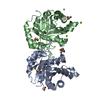


 PDBj
PDBj
 Assembly


 Mass: 118.174 Da / Num. of mol.: 5 / Source method: obtained synthetically / Formula: C6H14O2 / Comment: precipitant*YM
 Mass: 118.174 Da / Num. of mol.: 1 / Source method: obtained synthetically / Formula: C6H14O2 / Comment: precipitant*YM
 Mass: 18.015 Da / Num. of mol.: 250 / Source method: isolated from a natural source / Formula: H2O
 Sample preparation
 / Beamline: PROXIMA 1 / Wavelength: 0.885 Å
 Processing