 Movie
Movie Controller

[English] 日本語
 Yorodumi
- PDB-1aw1: TRIOSEPHOSPHATE ISOMERASE OF VIBRIO MARINUS COMPLEXED WITH 2-PHOS... -
+ Open data

- Basic information
| Entry | Database: PDB / ID: 1aw1 | ||||||
|---|---|---|---|---|---|---|---|
| Title | TRIOSEPHOSPHATE ISOMERASE OF VIBRIO MARINUS COMPLEXED WITH 2-PHOSPHOGLYCOLATE | ||||||
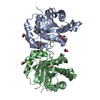
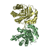
 Components Components | TRIOSEPHOSPHATE ISOMERASE | ||||||
 Keywords Keywords | ISOMERASE / PSYCHROPHILIC / VIBRIO MARINUS | ||||||
| Function / homology |  Function and homology information Function and homology informationtriose-phosphate isomerase / triose-phosphate isomerase activity / glyceraldehyde-3-phosphate biosynthetic process / glycerol catabolic process / gluconeogenesis / glycolytic process / cytosol Similarity search - Function | ||||||
| Biological species |  Moritella marina (bacteria) Moritella marina (bacteria) | ||||||
| Method |  X-RAY DIFFRACTION / MOLECULAR REPLACEMENT / Resolution: 2.7 Å X-RAY DIFFRACTION / MOLECULAR REPLACEMENT / Resolution: 2.7 Å | ||||||
 Authors Authors | Maes, D. / Zeelen, J.P. / Wierenga, R.K. | ||||||
 Citation Citation | Journal: J.Biol.Chem. / Year: 1998 Title: Triose-phosphate isomerase (TIM) of the psychrophilic bacterium Vibrio marinus. Kinetic and structural properties. Authors: Alvarez, M. / Zeelen, J.P. / Mainfroid, V. / Rentier-Delrue, F. / Martial, J.A. / Wyns, L. / Wierenga, R.K. / Maes, D. | ||||||
| History |
|
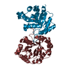
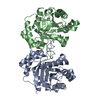
- Structure visualization
| Structure viewer | Molecule: MolmilJmol/JSmol |
|---|
- Downloads & links
-Download
| PDBx/mmCIF format | 1aw1.cif.gz | 367.8 KB | Display | PDBx/mmCIF format |
|---|---|---|---|---|
| PDB format | pdb1aw1.ent.gz | 304.4 KB | Display | PDB format |
| PDBx/mmJSON format | 1aw1.json.gz | Tree view | PDBx/mmJSON format | |
| Others |  Other downloads Other downloads |
-Validation report
| Arichive directory | https://data.pdbj.org/pub/pdb/validation_reports/aw/1aw1ftp://data.pdbj.org/pub/pdb/validation_reports/aw/1aw1 | HTTPS FTP |
|---|
-Related structure data
| Related structure data |  1aw2C 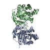 1treS S: Starting model for refinement C: citing same article ( |
|---|---|
| Similar structure data |

-Links
 PDBj
PDBj
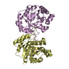
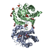
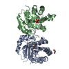
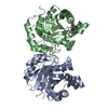
- Assembly
| Deposited unit | 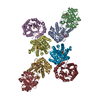
| ||||||||||||||||
|---|---|---|---|---|---|---|---|---|---|---|---|---|---|---|---|---|---|
| 1 | 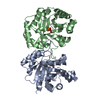
| ||||||||||||||||
| 2 | 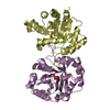
| ||||||||||||||||
| 3 | 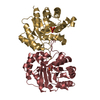
| ||||||||||||||||
| 4 | 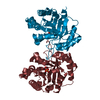
| ||||||||||||||||
| Unit cell |
| ||||||||||||||||
| Noncrystallographic symmetry (NCS) | NCS oper:
|
-Components
| #1: Protein | Mass: 26766.199 Da / Num. of mol.: 8 Source method: isolated from a genetically manipulated source Details: COMPLEX WITH 2-PHOSPHOGLYCOLATE / Source: (gene. exp.) Moritella marina (bacteria) / Production host: #2: Chemical | ChemComp-PGA / 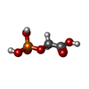  Mass: 156.031 Da / Num. of mol.: 8 Mass: 156.031 Da / Num. of mol.: 8Source method: isolated from a genetically manipulated source Formula: C2H5O6P #3: Water | ChemComp-HOH / |  Mass: 18.015 Da / Num. of mol.: 308 / Source method: isolated from a natural source / Formula: H2O Mass: 18.015 Da / Num. of mol.: 308 / Source method: isolated from a natural source / Formula: H2O |
|---|
-Experimental details
-Experiment
| Experiment | Method: X-RAY DIFFRACTION / Number of used crystals: 1 |
|---|
- Sample preparation
| Crystal | Density Matthews: 2.5 Å3/Da / Density % sol: 52 % | ||||||||||||||||||||||||||||||||||||||||||||||||
|---|---|---|---|---|---|---|---|---|---|---|---|---|---|---|---|---|---|---|---|---|---|---|---|---|---|---|---|---|---|---|---|---|---|---|---|---|---|---|---|---|---|---|---|---|---|---|---|---|---|
| Crystal grow | pH: 7.5 Details: 100MM TRIETHANOLAMINE/HCL 2.OM AMMONIUM SULFATE, 1MM DTT, EDTA, NAN3 20MM PGA PH 7.5 | ||||||||||||||||||||||||||||||||||||||||||||||||
| Crystal grow | *PLUS Temperature: 4 ℃ / Method: vapor diffusion, hanging drop | ||||||||||||||||||||||||||||||||||||||||||||||||
| Components of the solutions | *PLUS
|
-Data collection
| Diffraction | Mean temperature: 298 K |
|---|---|
| Diffraction source | Source: ROTATING ANODE / Type: ENRAF-NONIUS FR571 / Wavelength: 1.5418 |
| Detector | Type: MARRESEARCH / Detector: IMAGE PLATE / Date: Nov 1, 1996 / Details: DOUBLE FOCUSSING MIRRORS |
| Radiation | Monochromatic (M) / Laue (L): M / Scattering type: x-ray |
| Radiation wavelength | Wavelength: 1.5418 Å / Relative weight: 1 |
| Reflection | Resolution: 2.7→25 Å / Num. obs: 59692 / % possible obs: 99.9 % / Redundancy: 3.7 % / Biso Wilson estimate: 34 Å2 / Rmerge(I) obs: 0.084 / Net I/σ(I): 14 |
| Reflection shell | Resolution: 2.7→2.75 Å / Redundancy: 3 % / Rmerge(I) obs: 0.198 / Mean I/σ(I) obs: 7 / % possible all: 98.2 |
| Reflection | *PLUS Num. measured all: 332463 |
| Reflection shell | *PLUS % possible obs: 98.2 % |
- Processing
| Software |
| ||||||||||||||||||||||||||||||||||||||||||||||||||||||||||||
|---|---|---|---|---|---|---|---|---|---|---|---|---|---|---|---|---|---|---|---|---|---|---|---|---|---|---|---|---|---|---|---|---|---|---|---|---|---|---|---|---|---|---|---|---|---|---|---|---|---|---|---|---|---|---|---|---|---|---|---|---|---|
| Refinement | Method to determine structure: MOLECULAR REPLACEMENT Starting model: PDB ENTRY 1TRE Resolution: 2.7→8 Å / Data cutoff high absF: 10000000 / Data cutoff low absF: 0.01 / Cross valid method: THROUGHOUT / σ(F): 0
| ||||||||||||||||||||||||||||||||||||||||||||||||||||||||||||
| Displacement parameters | Biso mean: 14 Å2 | ||||||||||||||||||||||||||||||||||||||||||||||||||||||||||||
| Refinement step | Cycle: LAST / Resolution: 2.7→8 Å
| ||||||||||||||||||||||||||||||||||||||||||||||||||||||||||||
| Refine LS restraints |
| ||||||||||||||||||||||||||||||||||||||||||||||||||||||||||||
| Refine LS restraints NCS | NCS model details: CONSTRAINTS | ||||||||||||||||||||||||||||||||||||||||||||||||||||||||||||
| LS refinement shell | Resolution: 2.7→2.82 Å / Total num. of bins used: 8
| ||||||||||||||||||||||||||||||||||||||||||||||||||||||||||||
| Xplor file |
| ||||||||||||||||||||||||||||||||||||||||||||||||||||||||||||
| Software | *PLUS Name: X-PLOR / Version: 3.8 / Classification: refinement | ||||||||||||||||||||||||||||||||||||||||||||||||||||||||||||
| Refinement | *PLUS | ||||||||||||||||||||||||||||||||||||||||||||||||||||||||||||
| Solvent computation | *PLUS | ||||||||||||||||||||||||||||||||||||||||||||||||||||||||||||
| Displacement parameters | *PLUS | ||||||||||||||||||||||||||||||||||||||||||||||||||||||||||||
| Refine LS restraints | *PLUS
| ||||||||||||||||||||||||||||||||||||||||||||||||||||||||||||
| LS refinement shell | *PLUS Rfactor obs: 0.25 |
