 Movie
Movie Controller

[English] 日本語
 Yorodumi
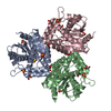
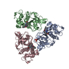
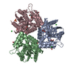
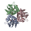
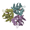
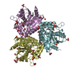
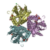
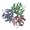
- PDB-6x3j: Crystal structure of streptogramin A acetyltransferase VatA from ... -
+ Open data

- Basic information
| Entry | Database: PDB / ID: 6x3j | ||||||
|---|---|---|---|---|---|---|---|
| Title | Crystal structure of streptogramin A acetyltransferase VatA from Staphylococcus aureus in complex with streptogramin analog F0224 (46) | ||||||
 Components Components | Virginiamycin A acetyltransferase,VatA | ||||||
 Keywords Keywords | TRANSFERASE / Acetyltransferase | ||||||
| Function / homology |  Function and homology information Function and homology informationacyltransferase activity / Transferases; Acyltransferases; Transferring groups other than aminoacyl groups / response to antibiotic Similarity search - Function | ||||||
| Biological species |   Staphylococcus aureus (bacteria) Staphylococcus aureus (bacteria) | ||||||
| Method |  X-RAY DIFFRACTION / SYNCHROTRON / MOLECULAR REPLACEMENT / Resolution: 2.7 Å X-RAY DIFFRACTION / SYNCHROTRON / MOLECULAR REPLACEMENT / Resolution: 2.7 Å | ||||||
 Authors Authors | Chaires, H.A. / Fraser, J.S. | ||||||
| Funding support |  United States, 1items United States, 1items
| ||||||
 Citation Citation | Journal: Nature / Year: 2020 Title: Synthetic group A streptogramin antibiotics that overcome Vat resistance. Authors: Qi Li / Jenna Pellegrino / D John Lee / Arthur A Tran / Hector A Chaires / Ruoxi Wang / Jesslyn E Park / Kaijie Ji / David Chow / Na Zhang / Axel F Brilot / Justin T Biel / Gydo van Zundert ...Authors: Qi Li / Jenna Pellegrino / D John Lee / Arthur A Tran / Hector A Chaires / Ruoxi Wang / Jesslyn E Park / Kaijie Ji / David Chow / Na Zhang / Axel F Brilot / Justin T Biel / Gydo van Zundert / Kenneth Borrelli / Dean Shinabarger / Cindy Wolfe / Beverly Murray / Matthew P Jacobson / Estelle Mühle / Olivier Chesneau / James S Fraser / Ian B Seiple /   Abstract: Natural products serve as chemical blueprints for most antibiotics in clinical use. The evolutionary process by which these molecules arise is inherently accompanied by the co-evolution of resistance ...Natural products serve as chemical blueprints for most antibiotics in clinical use. The evolutionary process by which these molecules arise is inherently accompanied by the co-evolution of resistance mechanisms that shorten the clinical lifetime of any given class of antibiotics. Virginiamycin acetyltransferase (Vat) enzymes are resistance proteins that provide protection against streptogramins, potent antibiotics against Gram-positive bacteria that inhibit the bacterial ribosome. Owing to the challenge of selectively modifying the chemically complex, 23-membered macrocyclic scaffold of group A streptogramins, analogues that overcome the resistance conferred by Vat enzymes have not been previously developed. Here we report the design, synthesis, and antibacterial evaluation of group A streptogramin antibiotics with extensive structural variability. Using cryo-electron microscopy and forcefield-based refinement, we characterize the binding of eight analogues to the bacterial ribosome at high resolution, revealing binding interactions that extend into the peptidyl tRNA-binding site and towards synergistic binders that occupy the nascent peptide exit tunnel. One of these analogues has excellent activity against several streptogramin-resistant strains of Staphylococcus aureus, exhibits decreased rates of acetylation in vitro, and is effective at lowering bacterial load in a mouse model of infection. Our results demonstrate that the combination of rational design and modular chemical synthesis can revitalize classes of antibiotics that are limited by naturally arising resistance mechanisms. | ||||||
| History |
|
- Structure visualization
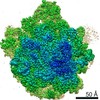
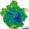
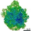
| Structure viewer | Molecule: MolmilJmol/JSmol |
|---|
- Downloads & links
-Download
| PDBx/mmCIF format | 6x3j.cif.gz | 788.3 KB | Display | PDBx/mmCIF format |
|---|---|---|---|---|
| PDB format | pdb6x3j.ent.gz | 647.4 KB | Display | PDB format |
| PDBx/mmJSON format | 6x3j.json.gz | Tree view | PDBx/mmJSON format | |
| Others |  Other downloads Other downloads |
-Validation report
| Arichive directory | https://data.pdbj.org/pub/pdb/validation_reports/x3/6x3jftp://data.pdbj.org/pub/pdb/validation_reports/x3/6x3j | HTTPS FTP |
|---|
-Related structure data
| Related structure data |  6pc5C  6pc6C  6pc7C  6pc8C 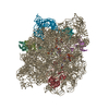 6pchC 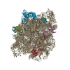 6pcqC 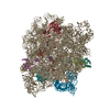 6pcrC  6pcsC  6pctC  6wyvC  6x3cC  4hurS C: citing same article ( S: Starting model for refinement |
|---|---|
| Similar structure data |


-Links
 PDBj
PDBj


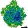
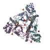
- Assembly
| Deposited unit | 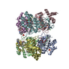
| ||||||||||||
|---|---|---|---|---|---|---|---|---|---|---|---|---|---|
| 1 | 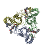
| ||||||||||||
| 2 | 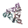
| ||||||||||||
| Unit cell |
|
-Components
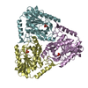
-Protein , 1 types, 6 molecules ABCDEF
| #1: Protein | Mass: 23814.494 Da / Num. of mol.: 6 Source method: isolated from a genetically manipulated source Source: (gene. exp.) Staphylococcus aureus (bacteria) / Gene: vat / Production host: References: UniProt: P26839, Transferases; Acyltransferases; Transferring groups other than aminoacyl groups |
|---|
-Non-polymers , 6 types, 235 molecules 


| #2: Chemical | ChemComp-O7V / (  Mass: 717.783 Da / Num. of mol.: 6 / Source method: obtained synthetically / Formula: C38H44FN5O8 / Feature type: SUBJECT OF INVESTIGATION Mass: 717.783 Da / Num. of mol.: 6 / Source method: obtained synthetically / Formula: C38H44FN5O8 / Feature type: SUBJECT OF INVESTIGATION#3: Chemical |  Mass: 94.971 Da / Num. of mol.: 2 / Source method: obtained synthetically / Formula: PO4 Mass: 94.971 Da / Num. of mol.: 2 / Source method: obtained synthetically / Formula: PO4#4: Chemical | ChemComp-SXA /  Mass: 400.385 Da / Num. of mol.: 6 / Source method: obtained synthetically / Formula: C13H25N2O8PS Mass: 400.385 Da / Num. of mol.: 6 / Source method: obtained synthetically / Formula: C13H25N2O8PS#5: Chemical | ChemComp-CL /  Mass: 35.453 Da / Num. of mol.: 11 / Source method: obtained synthetically / Formula: Cl Mass: 35.453 Da / Num. of mol.: 11 / Source method: obtained synthetically / Formula: Cl#6: Chemical |  Mass: 24.305 Da / Num. of mol.: 2 / Source method: obtained synthetically / Formula: Mg Mass: 24.305 Da / Num. of mol.: 2 / Source method: obtained synthetically / Formula: Mg#7: Water | ChemComp-HOH / | Mass: 18.015 Da / Num. of mol.: 208 / Source method: isolated from a natural source / Formula: H2O |
|---|
-Details
| Has ligand of interest | Y |
|---|
-Experimental details
-Experiment
| Experiment | Method: X-RAY DIFFRACTION / Number of used crystals: 1 |
|---|
- Sample preparation
| Crystal | Density Matthews: 3.3 Å3/Da / Density % sol: 62.71 % |
|---|---|
| Crystal grow | Temperature: 293 K / Method: vapor diffusion, hanging drop / Details: 1 M LiCl, 0.1 M BICINE pH 9, and 10 %w/v PEG 6K |
-Data collection
| Diffraction | Mean temperature: 92 K / Serial crystal experiment: N |
|---|---|
| Diffraction source | Source: SYNCHROTRON / Site: ALS / Beamline: 8.3.1 / Wavelength: 1.11583 Å |
| Detector | Type: DECTRIS PILATUS3 6M / Detector: PIXEL / Date: Aug 30, 2019 |
| Radiation | Protocol: SINGLE WAVELENGTH / Monochromatic (M) / Laue (L): M / Scattering type: x-ray |
| Radiation wavelength | Wavelength: 1.11583 Å / Relative weight: 1 |
| Reflection | Resolution: 2.7→86.51 Å / Num. obs: 55239 / % possible obs: 99.96 % / Redundancy: 29.6 % / Biso Wilson estimate: 57.72 Å2 / CC1/2: 0.995 / CC star: 0.999 / Net I/σ(I): 9.41 |
| Reflection shell | Resolution: 2.7→2.797 Å / Num. unique obs: 5440 / CC1/2: 0.404 / CC star: 0.758 / % possible all: 99.96 |
- Processing
| Software |
| ||||||||||||||||||||||||
|---|---|---|---|---|---|---|---|---|---|---|---|---|---|---|---|---|---|---|---|---|---|---|---|---|---|
| Refinement | Method to determine structure: MOLECULAR REPLACEMENT Starting model: 4HUR Resolution: 2.7→86.51 Å / Cross valid method: FREE R-VALUE Stereochemistry target values: GeoStd + Monomer Library + CDL v1.2
| ||||||||||||||||||||||||
| Displacement parameters | Biso mean: 57.72 Å2 | ||||||||||||||||||||||||
| Refinement step | Cycle: LAST / Resolution: 2.7→86.51 Å
| ||||||||||||||||||||||||
| Refine LS restraints |
|
