 Movie
Movie Controller

[English] 日本語
 Yorodumi
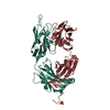
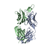
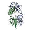
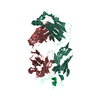
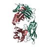
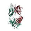
- PDB-6x1w: Structure of pHis Fab (SC56-2) in complex with pHis mimetic peptide -
+ Open data

- Basic information
| Entry | Database: PDB / ID: 6x1w | ||||||
|---|---|---|---|---|---|---|---|
| Title | Structure of pHis Fab (SC56-2) in complex with pHis mimetic peptide | ||||||
 Components Components |
| ||||||
 Keywords Keywords | IMMUNE SYSTEM / Anti-phosphohistidine antibody / post-translational modification | ||||||
| Function / homology | Immunoglobulins / Immunoglobulin-like / Sandwich / Mainly Beta Function and homology information Function and homology information | ||||||
| Biological species |  synthetic construct (others) | ||||||
| Method |  X-RAY DIFFRACTION / SYNCHROTRON / MOLECULAR REPLACEMENT / Resolution: 1.95 Å X-RAY DIFFRACTION / SYNCHROTRON / MOLECULAR REPLACEMENT / Resolution: 1.95 Å | ||||||
 Authors Authors | Kalagiri, R. / Stanfield, R. / Wilson, I.A. / Hunter, T. | ||||||
| Funding support |  United States, 1items United States, 1items
| ||||||
 Citation Citation | Journal: Proc.Natl.Acad.Sci.USA / Year: 2021 Title: Structural basis for differential recognition of phosphohistidine-containing peptides by 1-pHis and 3-pHis monoclonal antibodies. Authors: Kalagiri, R. / Stanfield, R.L. / Meisenhelder, J. / La Clair, J.J. / Fuhs, S.R. / Wilson, I.A. / Hunter, T. | ||||||
| History |
|
- Structure visualization
| Structure viewer | Molecule: MolmilJmol/JSmol |
|---|
- Downloads & links
-Download
| PDBx/mmCIF format | 6x1w.cif.gz | 105.9 KB | Display | PDBx/mmCIF format |
|---|---|---|---|---|
| PDB format | pdb6x1w.ent.gz | 78.2 KB | Display | PDB format |
| PDBx/mmJSON format | 6x1w.json.gz | Tree view | PDBx/mmJSON format | |
| Others |  Other downloads Other downloads |
-Validation report
| Summary document | 6x1w_validation.pdf.gz | 445.3 KB | Display | wwPDB validaton report |
|---|---|---|---|---|
| Full document | 6x1w_full_validation.pdf.gz | 448.5 KB | Display | |
| Data in XML | 6x1w_validation.xml.gz | 21.2 KB | Display | |
| Data in CIF | 6x1w_validation.cif.gz | 31.2 KB | Display | |
| Arichive directory | https://data.pdbj.org/pub/pdb/validation_reports/x1/6x1wftp://data.pdbj.org/pub/pdb/validation_reports/x1/6x1w | HTTPS FTP |
-Related structure data
| Related structure data |  6x1sC 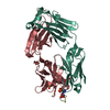 6x1tC  6x1uC  6x1vC  5dtfS S: Starting model for refinement C: citing same article ( |
|---|---|
| Similar structure data |


-Links
 PDBj
PDBj

- Assembly
| Deposited unit | 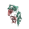
| ||||||||
|---|---|---|---|---|---|---|---|---|---|
| 1 |
| ||||||||
| Unit cell |
|
-Components
| #1: Antibody | Mass: 23539.363 Da / Num. of mol.: 1 Source method: isolated from a genetically manipulated source Source: (gene. exp.)  Homo sapiens (human) Homo sapiens (human) |
|---|---|
| #2: Antibody | Mass: 23576.783 Da / Num. of mol.: 1 Source method: isolated from a genetically manipulated source Source: (gene. exp.) Homo sapiens (human) |
| #3: Protein/peptide | Mass: 748.641 Da / Num. of mol.: 1 / Source method: obtained synthetically / Source: (synth.) synthetic construct (others) |
| #4: Water | ChemComp-HOH /  Mass: 18.015 Da / Num. of mol.: 345 / Source method: isolated from a natural source / Formula: H2O Mass: 18.015 Da / Num. of mol.: 345 / Source method: isolated from a natural source / Formula: H2O |
| Has ligand of interest | Y |
| Has protein modification | Y |
-Experimental details
-Experiment
| Experiment | Method: X-RAY DIFFRACTION / Number of used crystals: 1 |
|---|
- Sample preparation
| Crystal | Density Matthews: 3 Å3/Da / Density % sol: 58.99 % |
|---|---|
| Crystal grow | Temperature: 293 K / Method: vapor diffusion, sitting drop / pH: 6.5 / Details: 0.1 M Sodium cacodylate pH 6.5, 1 M Sodium citrate |
-Data collection
| Diffraction | Mean temperature: 100 K / Serial crystal experiment: N |
|---|---|
| Diffraction source | Source: SYNCHROTRON / Site: APS / Beamline: 23-ID-D / Wavelength: 1.0332 Å |
| Detector | Type: DECTRIS PILATUS3 S 6M / Detector: PIXEL / Date: Jun 10, 2017 |
| Radiation | Protocol: SINGLE WAVELENGTH / Monochromatic (M) / Laue (L): M / Scattering type: x-ray |
| Radiation wavelength | Wavelength: 1.0332 Å / Relative weight: 1 |
| Reflection | Resolution: 1.95→47.86 Å / Num. obs: 42281 / % possible obs: 99.1 % / Redundancy: 6.1 % / Biso Wilson estimate: 26.7 Å2 / CC1/2: 0.779 / Rmerge(I) obs: 0.152 / Rpim(I) all: 0.063 / Rrim(I) all: 0.165 / Net I/av σ(I): 10.6 / Net I/σ(I): 10.6 |
| Reflection shell | Resolution: 1.95→1.98 Å / Redundancy: 6.2 % / Rmerge(I) obs: 1.29 / Mean I/σ(I) obs: 1.1 / Num. unique obs: 2083 / CC1/2: 0.306 / Rpim(I) all: 0.538 / Rrim(I) all: 1.41 / % possible all: 99.8 |
- Processing
| Software |
| |||||||||||||||||||||||||||||||||||||||||||||||||||||||||||||||||||||||||||
|---|---|---|---|---|---|---|---|---|---|---|---|---|---|---|---|---|---|---|---|---|---|---|---|---|---|---|---|---|---|---|---|---|---|---|---|---|---|---|---|---|---|---|---|---|---|---|---|---|---|---|---|---|---|---|---|---|---|---|---|---|---|---|---|---|---|---|---|---|---|---|---|---|---|---|---|---|
| Refinement | Method to determine structure: MOLECULAR REPLACEMENT Starting model: 5dtf Resolution: 1.95→47.86 Å / Cor.coef. Fo:Fc: 0.957 / Cor.coef. Fo:Fc free: 0.937 / Cross valid method: THROUGHOUT / σ(F): 0 / ESU R: 0.149 / ESU R Free: 0.144 / Stereochemistry target values: MAXIMUM LIKELIHOOD Details: HYDROGENS HAVE BEEN ADDED IN THE RIDING POSITIONS U VALUES : REFINED INDIVIDUALLY
| |||||||||||||||||||||||||||||||||||||||||||||||||||||||||||||||||||||||||||
| Solvent computation | Ion probe radii: 0.8 Å / Shrinkage radii: 0.8 Å / VDW probe radii: 1.2 Å / Solvent model: MASK | |||||||||||||||||||||||||||||||||||||||||||||||||||||||||||||||||||||||||||
| Displacement parameters | Biso max: 130.85 Å2 / Biso mean: 30.809 Å2 / Biso min: 13.86 Å2
| |||||||||||||||||||||||||||||||||||||||||||||||||||||||||||||||||||||||||||
| Refinement step | Cycle: final / Resolution: 1.95→47.86 Å
| |||||||||||||||||||||||||||||||||||||||||||||||||||||||||||||||||||||||||||
| Refine LS restraints |
| |||||||||||||||||||||||||||||||||||||||||||||||||||||||||||||||||||||||||||
| LS refinement shell | Resolution: 1.951→2.002 Å / Total num. of bins used: 20
|
