 Movie
Movie Controller

[English] 日本語
 Yorodumi
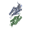
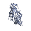
- PDB-6x1j: The homing endonuclease I-WcaI bound to its DNA recognition sequence -
+ Open data

- Basic information
| Entry | Database: PDB / ID: 6x1j | ||||||
|---|---|---|---|---|---|---|---|
| Title | The homing endonuclease I-WcaI bound to its DNA recognition sequence | ||||||
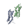
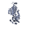
 Components Components |
| ||||||
 Keywords Keywords | HYDROLASE / intron-encoded DNA homing endonuclease / enzyme / protein-DNA interactions / isoschizomers | ||||||
| Function / homology |  Function and homology information Function and homology informationGroup II intron splicing / intron homing / mRNA cis splicing, via spliceosome / endonuclease activity / Hydrolases; Acting on ester bonds / mitochondrion Similarity search - Function | ||||||
| Biological species |  Wickerhamomyces canadensis (fungus) Wickerhamomyces canadensis (fungus) | ||||||
| Method |  X-RAY DIFFRACTION / SYNCHROTRON / MOLECULAR REPLACEMENT / molecular replacement / Resolution: 1.945 Å X-RAY DIFFRACTION / SYNCHROTRON / MOLECULAR REPLACEMENT / molecular replacement / Resolution: 1.945 Å | ||||||
 Authors Authors | Nawimanage, R. / Lohman, J.R. / Gimble, F.S. | ||||||
 Citation Citation | Journal: J.Mol.Biol. / Year: 2022 Title: Structure-Function Studies of Two Yeast Homing Endonucleases that Evolved to Cleave Identical Targets with Dissimilar Rates and Specificities. Authors: Nawimanage, R.R. / Yuan, Z. / Casares, M. / Joshi, R. / Lohman, J.R. / Gimble, F.S. | ||||||
| History |
|
- Structure visualization
| Structure viewer | Molecule: MolmilJmol/JSmol |
|---|
- Downloads & links
-Download
| PDBx/mmCIF format | 6x1j.cif.gz | 98 KB | Display | PDBx/mmCIF format |
|---|---|---|---|---|
| PDB format | pdb6x1j.ent.gz | 68 KB | Display | PDB format |
| PDBx/mmJSON format | 6x1j.json.gz | Tree view | PDBx/mmJSON format | |
| Others |  Other downloads Other downloads |
-Validation report
| Summary document | 6x1j_validation.pdf.gz | 442.1 KB | Display | wwPDB validaton report |
|---|---|---|---|---|
| Full document | 6x1j_full_validation.pdf.gz | 444.8 KB | Display | |
| Data in XML | 6x1j_validation.xml.gz | 13.5 KB | Display | |
| Data in CIF | 6x1j_validation.cif.gz | 19.1 KB | Display | |
| Arichive directory | https://data.pdbj.org/pub/pdb/validation_reports/x1/6x1jftp://data.pdbj.org/pub/pdb/validation_reports/x1/6x1j | HTTPS FTP |
-Related structure data
| Related structure data | 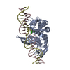 1r7mS S: Starting model for refinement |
|---|---|
| Similar structure data |


-Links
 PDBj
PDBj


- Assembly
| Deposited unit | 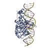
| ||||||||
|---|---|---|---|---|---|---|---|---|---|
| 1 |
| ||||||||
| Unit cell |
| ||||||||
| Components on special symmetry positions |
|
-Components
| #1: Protein | Mass: 27855.705 Da / Num. of mol.: 1 Source method: isolated from a genetically manipulated source Source: (gene. exp.) Wickerhamomyces canadensis (fungus) / Production host:  References: UniProt: Q34807, Hydrolases; Acting on ester bonds | ||||
|---|---|---|---|---|---|
| #2: DNA chain | Mass: 7741.026 Da / Num. of mol.: 1 / Source method: obtained synthetically / Source: (synth.) Wickerhamomyces canadensis (fungus) | ||||
| #3: DNA chain | Mass: 7615.906 Da / Num. of mol.: 1 / Source method: obtained synthetically / Source: (synth.) Wickerhamomyces canadensis (fungus) | ||||
| #4: Chemical | ChemComp-K /   Mass: 39.098 Da / Num. of mol.: 4 / Source method: obtained synthetically / Formula: K Mass: 39.098 Da / Num. of mol.: 4 / Source method: obtained synthetically / Formula: K#5: Water | ChemComp-HOH / |  Mass: 18.015 Da / Num. of mol.: 140 / Source method: isolated from a natural source / Formula: H2O Mass: 18.015 Da / Num. of mol.: 140 / Source method: isolated from a natural source / Formula: H2OHas ligand of interest | N | |
-Experimental details
-Experiment
| Experiment | Method: X-RAY DIFFRACTION / Number of used crystals: 1 |
|---|
- Sample preparation
| Crystal | Density Matthews: 2.74 Å3/Da / Density % sol: 55.09 % / Description: Needles |
|---|---|
| Crystal grow | Temperature: 298 K / Method: vapor diffusion, hanging drop / pH: 8.5 Details: 21% PEG 3350, 0.1M Tris-HCl, 0.2M Lithium sulfate, 2 mM DTT |
-Data collection
| Diffraction | Mean temperature: 100 K / Serial crystal experiment: N |
|---|---|
| Diffraction source | Source: SYNCHROTRON / Site: APS  / Beamline: 21-ID-F / Wavelength: 0.97872 Å / Beamline: 21-ID-F / Wavelength: 0.97872 Å |
| Detector | Type: RAYONIX MX300HE / Detector: CCD / Date: Feb 17, 2016 |
| Radiation | Monochromator: C(111) / Protocol: SINGLE WAVELENGTH / Monochromatic (M) / Laue (L): M / Scattering type: x-ray |
| Radiation wavelength | Wavelength: 0.97872 Å / Relative weight: 1 |
| Reflection | Resolution: 1.945→30 Å / Num. obs: 33735 / % possible obs: 99.8 % / Redundancy: 3.6 % / Biso Wilson estimate: 33.3 Å2 / CC1/2: 0.964 / CC star: 0.991 / Rpim(I) all: 0.104 / Rrim(I) all: 0.198 / Χ2: 1.063 / Net I/σ(I): 6.5 |
| Reflection shell | Resolution: 1.945→2.02 Å / Redundancy: 3.3 % / Rmerge(I) obs: 0.72 / Mean I/σ(I) obs: 1.6 / Num. unique obs: 3306 / CC1/2: 0.807 / CC star: 0.945 / Rpim(I) all: 0.461 / Rrim(I) all: 0.858 / Χ2: 0.882 / % possible all: 99.6 |
-Phasing
| Phasing | Method: molecular replacement | |||||||||
|---|---|---|---|---|---|---|---|---|---|---|
| Phasing MR | Model details: Phaser MODE: MR_AUTO
|
- Processing
| Software |
| ||||||||||||||||||||||||||||||||||||||||||||||||||||||||||||
|---|---|---|---|---|---|---|---|---|---|---|---|---|---|---|---|---|---|---|---|---|---|---|---|---|---|---|---|---|---|---|---|---|---|---|---|---|---|---|---|---|---|---|---|---|---|---|---|---|---|---|---|---|---|---|---|---|---|---|---|---|---|
| Refinement | Method to determine structure: MOLECULAR REPLACEMENT Starting model: 1R7M Resolution: 1.945→29.88 Å / Cor.coef. Fo:Fc: 0.954 / Cor.coef. Fo:Fc free: 0.936 / SU B: 8.699 / SU ML: 0.212 / Cross valid method: THROUGHOUT / σ(F): 0 / ESU R: 0.181 / ESU R Free: 0.163 / Stereochemistry target values: MAXIMUM LIKELIHOOD Details: HYDROGENS HAVE BEEN ADDED IN THE RIDING POSITIONS U VALUES : REFINED INDIVIDUALLY
| ||||||||||||||||||||||||||||||||||||||||||||||||||||||||||||
| Solvent computation | Ion probe radii: 0.8 Å / Shrinkage radii: 0.8 Å / VDW probe radii: 1.2 Å / Solvent model: MASK | ||||||||||||||||||||||||||||||||||||||||||||||||||||||||||||
| Displacement parameters | Biso max: 127.33 Å2 / Biso mean: 37.752 Å2 / Biso min: 12.59 Å2
| ||||||||||||||||||||||||||||||||||||||||||||||||||||||||||||
| Refinement step | Cycle: final / Resolution: 1.945→29.88 Å
| ||||||||||||||||||||||||||||||||||||||||||||||||||||||||||||
| Refine LS restraints |
| ||||||||||||||||||||||||||||||||||||||||||||||||||||||||||||
| LS refinement shell | Resolution: 1.9451→1.996 Å / Rfactor Rfree error: 0
|
