 Movie
Movie Controller

[English] 日本語
 Yorodumi
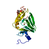
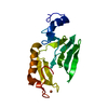
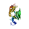
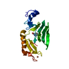
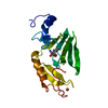
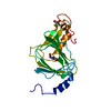
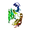
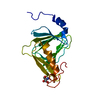
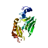
- PDB-4l2n: Understanding Extradiol Dioxygenase Mechanism in NAD+ Biosynthesi... -
+ Open data

- Basic information
| Entry | Database: PDB / ID: 4l2n | ||||||
|---|---|---|---|---|---|---|---|
| Title | Understanding Extradiol Dioxygenase Mechanism in NAD+ Biosynthesis by Viewing Catalytic Intermediates - ligand-free structure | ||||||
 Components Components | 3-hydroxyanthranilate 3,4-dioxygenase | ||||||
 Keywords Keywords | OXIDOREDUCTASE / bi-cupin iron-binding / dioxygenase | ||||||
| Function / homology |  Function and homology information Function and homology information3-hydroxyanthranilate 3,4-dioxygenase / 3-hydroxyanthranilate 3,4-dioxygenase activity / : / L-tryptophan catabolic process / quinolinate biosynthetic process / NAD+ biosynthetic process / ferrous iron binding Similarity search - Function | ||||||
| Biological species |  Cupriavidus metallidurans (bacteria) Cupriavidus metallidurans (bacteria) | ||||||
| Method |  X-RAY DIFFRACTION / SYNCHROTRON / MOLECULAR REPLACEMENT / Resolution: 1.74 Å X-RAY DIFFRACTION / SYNCHROTRON / MOLECULAR REPLACEMENT / Resolution: 1.74 Å | ||||||
 Authors Authors | Liu, F. / Liu, A. | ||||||
 Citation Citation | Journal: J.Biol.Chem. / Year: 2015 Title: An Iron Reservoir to the Catalytic Metal: THE RUBREDOXIN IRON IN AN EXTRADIOL DIOXYGENASE. Authors: Liu, F. / Geng, J. / Gumpper, R.H. / Barman, A. / Davis, I. / Ozarowski, A. / Hamelberg, D. / Liu, A. | ||||||
| History |
|
- Structure visualization
| Structure viewer | Molecule: MolmilJmol/JSmol |
|---|
- Downloads & links
-Download
| PDBx/mmCIF format | 4l2n.cif.gz | 53.3 KB | Display | PDBx/mmCIF format |
|---|---|---|---|---|
| PDB format | pdb4l2n.ent.gz | 36.5 KB | Display | PDB format |
| PDBx/mmJSON format | 4l2n.json.gz | Tree view | PDBx/mmJSON format | |
| Others |  Other downloads Other downloads |
-Validation report
| Arichive directory | https://data.pdbj.org/pub/pdb/validation_reports/l2/4l2nftp://data.pdbj.org/pub/pdb/validation_reports/l2/4l2n | HTTPS FTP |
|---|
-Related structure data
| Related structure data |  4hsjC  4hvoC  4hvqC  1yfuS  4l2p 4l2q 4l2r 4l2s 4l2t 4l2u 4l2v S: Starting model for refinement C: citing same article ( |
|---|---|
| Similar structure data |

-Links
 PDBj
PDBj


- Assembly
| Deposited unit | 
| ||||||||
|---|---|---|---|---|---|---|---|---|---|
| 1 |
| ||||||||
| 2 | 
| ||||||||
| Unit cell |
|
-Components
| #1: Protein | Mass: 20056.635 Da / Num. of mol.: 1 Source method: isolated from a genetically manipulated source Source: (gene. exp.) Cupriavidus metallidurans (bacteria) / Strain: CH34 / ATCC 43123 / DSM 2839 / Gene: Cupriavidus metallidurans, nbaC, Rmet_5193 / Production host: References: UniProt: Q1LCS4, 3-hydroxyanthranilate 3,4-dioxygenase | ||||||
|---|---|---|---|---|---|---|---|
| #2: Chemical |   Mass: 55.845 Da / Num. of mol.: 2 / Source method: obtained synthetically / Formula: Fe Mass: 55.845 Da / Num. of mol.: 2 / Source method: obtained synthetically / Formula: Fe#3: Chemical | ChemComp-CL / |   Mass: 35.453 Da / Num. of mol.: 1 / Source method: obtained synthetically / Formula: Cl Mass: 35.453 Da / Num. of mol.: 1 / Source method: obtained synthetically / Formula: Cl#4: Chemical | ChemComp-TRS / |   Mass: 122.143 Da / Num. of mol.: 1 / Source method: obtained synthetically / Formula: C4H12NO3 / Comment: pH buffer*YM Mass: 122.143 Da / Num. of mol.: 1 / Source method: obtained synthetically / Formula: C4H12NO3 / Comment: pH buffer*YM#5: Water | ChemComp-HOH / |  Mass: 18.015 Da / Num. of mol.: 117 / Source method: isolated from a natural source / Formula: H2O Mass: 18.015 Da / Num. of mol.: 117 / Source method: isolated from a natural source / Formula: H2O |
-Experimental details
-Experiment
| Experiment | Method: X-RAY DIFFRACTION / Number of used crystals: 1 |
|---|
- Sample preparation
| Crystal | Density Matthews: 2.83 Å3/Da / Density % sol: 56.6 % |
|---|---|
| Crystal grow | Temperature: 291 K / Method: vapor diffusion, hanging drop / pH: 8.5 Details: PEG 8000, 0.1M Tris-HCl, 200 mM MgCl2 , pH 8.5, VAPOR DIFFUSION, HANGING DROP, temperature 291K |
-Data collection
| Diffraction | Mean temperature: 100 K |
|---|---|
| Diffraction source | Source: SYNCHROTRON / Site: APS  / Beamline: 22-ID / Wavelength: 1 Å / Beamline: 22-ID / Wavelength: 1 Å |
| Detector | Type: MARMOSAIC 300 mm CCD / Detector: CCD / Date: Oct 24, 2012 |
| Radiation | Protocol: SINGLE WAVELENGTH / Monochromatic (M) / Laue (L): M / Scattering type: x-ray |
| Radiation wavelength | Wavelength: 1 Å / Relative weight: 1 |
| Reflection | Resolution: 1.74→50 Å / Num. all: 25017 / Num. obs: 24242 / % possible obs: 96.9 % / Observed criterion σ(F): 0 |
| Reflection shell | Resolution: 1.75→1.78 Å / % possible all: 76.8 |
- Processing
| Software |
| ||||||||||||||||||||||||||||||||||||
|---|---|---|---|---|---|---|---|---|---|---|---|---|---|---|---|---|---|---|---|---|---|---|---|---|---|---|---|---|---|---|---|---|---|---|---|---|---|
| Refinement | Method to determine structure: MOLECULAR REPLACEMENT Starting model: 1YFU Resolution: 1.74→34.129 Å / Occupancy max: 1 / Occupancy min: 1 / SU ML: 0.13 / Cross valid method: THROUGHOUT / σ(F): 1.34 / Phase error: 24.4 / Stereochemistry target values: MAXIMUM LIKELIHOOD
| ||||||||||||||||||||||||||||||||||||
| Solvent computation | Shrinkage radii: 0.73 Å / VDW probe radii: 1 Å / Solvent model: FLAT BULK SOLVENT MODEL / Bsol: 43.94 Å2 / ksol: 0.403 e/Å3 | ||||||||||||||||||||||||||||||||||||
| Displacement parameters | Biso max: 89.41 Å2 / Biso mean: 32.4994 Å2 / Biso min: 17.09 Å2
| ||||||||||||||||||||||||||||||||||||
| Refinement step | Cycle: LAST / Resolution: 1.74→34.129 Å
| ||||||||||||||||||||||||||||||||||||
| Refine LS restraints |
|
