 Movie
Movie Controller

[English] 日本語
 Yorodumi
- PDB-6wyn: Transition metal inhibition and structural refinement of the M. t... -
+ Open data

- Basic information
| Entry | Database: PDB / ID: 6wyn | ||||||
|---|---|---|---|---|---|---|---|
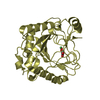
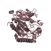
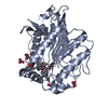
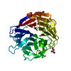
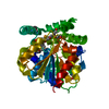
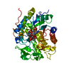
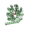
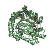
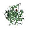
| Title | Transition metal inhibition and structural refinement of the M. tuberculosis esterase, Rv0045c | ||||||
 Components Components | Possible hydrolase | ||||||
 Keywords Keywords | HYDROLASE / serine esterase / allosteric regulation / conformational change | ||||||
| Function / homology | : / carboxylesterase / carboxylesterase activity / Alpha/beta hydrolase family / Alpha/beta hydrolase fold-1 / Alpha/Beta hydrolase fold / hydrolase activity / Esterase Rv0045c Function and homology information Function and homology information | ||||||
| Biological species |   Mycobacterium tuberculosis (bacteria) Mycobacterium tuberculosis (bacteria) | ||||||
| Method |  X-RAY DIFFRACTION / SYNCHROTRON / MOLECULAR REPLACEMENT / molecular replacement / Resolution: 1.81 Å X-RAY DIFFRACTION / SYNCHROTRON / MOLECULAR REPLACEMENT / molecular replacement / Resolution: 1.81 Å | ||||||
 Authors Authors | Macbeth, M.R. / Johnson, R.J. / Hoops, G.C. | ||||||
| Funding support |  United States, 1items United States, 1items
| ||||||
 Citation Citation | Journal: Protein Sci. / Year: 2021 Title: Transition metal cation inhibition of Mycobacterium tuberculosis esterase RV0045C. Authors: Bowles, I.E. / Pool, E.H. / Lancaster, B.S. / Lawson, E.K. / Savas, C.P. / Kartje, Z.J. / Severinac, L. / Cho, D.H. / Macbeth, M.R. / Johnson, R.J. / Hoops, G.C. | ||||||
| History |
|
- Structure visualization
| Structure viewer | Molecule: MolmilJmol/JSmol |
|---|
- Downloads & links
-Download
| PDBx/mmCIF format | 6wyn.cif.gz | 75.9 KB | Display | PDBx/mmCIF format |
|---|---|---|---|---|
| PDB format | pdb6wyn.ent.gz | 54.3 KB | Display | PDB format |
| PDBx/mmJSON format | 6wyn.json.gz | Tree view | PDBx/mmJSON format | |
| Others |  Other downloads Other downloads |
-Validation report
| Summary document | 6wyn_validation.pdf.gz | 430.9 KB | Display | wwPDB validaton report |
|---|---|---|---|---|
| Full document | 6wyn_full_validation.pdf.gz | 432.7 KB | Display | |
| Data in XML | 6wyn_validation.xml.gz | 14.4 KB | Display | |
| Data in CIF | 6wyn_validation.cif.gz | 21.1 KB | Display | |
| Arichive directory | https://data.pdbj.org/pub/pdb/validation_reports/wy/6wynftp://data.pdbj.org/pub/pdb/validation_reports/wy/6wyn | HTTPS FTP |
-Related structure data
| Related structure data |  6wymC  3p2mS C: citing same article ( S: Starting model for refinement |
|---|---|
| Similar structure data |

-Links
 PDBj
PDBj

- Assembly
| Deposited unit | 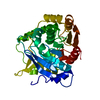
| ||||||||
|---|---|---|---|---|---|---|---|---|---|
| 1 |
| ||||||||
| Unit cell |
| ||||||||
| Components on special symmetry positions |
|
-Components
| #1: Protein | Mass: 35589.789 Da / Num. of mol.: 1 Source method: isolated from a genetically manipulated source Source: (gene. exp.) Mycobacterium tuberculosis (strain ATCC 25618 / H37Rv) (bacteria)Strain: ATCC 25618 / H37Rv / Gene: Rv0045c / Plasmid: pET-28 / Details (production host): KanR / Production host: |
|---|---|
| #2: Chemical | ChemComp-CL /   Mass: 35.453 Da / Num. of mol.: 1 / Source method: obtained synthetically / Formula: Cl Mass: 35.453 Da / Num. of mol.: 1 / Source method: obtained synthetically / Formula: Cl |
| #3: Water | ChemComp-HOH /  Mass: 18.015 Da / Num. of mol.: 212 / Source method: isolated from a natural source / Formula: H2O Mass: 18.015 Da / Num. of mol.: 212 / Source method: isolated from a natural source / Formula: H2O |
| Has ligand of interest | N |
-Experimental details
-Experiment
| Experiment | Method: X-RAY DIFFRACTION / Number of used crystals: 1 |
|---|
- Sample preparation
| Crystal | Density Matthews: 3.39 Å3/Da / Density % sol: 63.73 % |
|---|---|
| Crystal grow | Temperature: 294 K / Method: vapor diffusion, sitting drop / pH: 7.4 Details: 0.2 M MgCl2, 0.1 M Imidazole pH 7.4, 18% PEG 4000, 0.01 M spermidine, 1x10^-7 M ZnCl2 |
-Data collection
| Diffraction | Mean temperature: 100 K / Serial crystal experiment: N | ||||||||||||||||||||||||||||||
|---|---|---|---|---|---|---|---|---|---|---|---|---|---|---|---|---|---|---|---|---|---|---|---|---|---|---|---|---|---|---|---|
| Diffraction source | Source: SYNCHROTRON / Site: APS / Beamline: 31-ID / Wavelength: 1.279 Å | ||||||||||||||||||||||||||||||
| Detector | Type: DECTRIS PILATUS3 S 6M / Detector: PIXEL / Date: Jul 18, 2018 | ||||||||||||||||||||||||||||||
| Radiation | Monochromator: Kohzu HLD-4 Double Crystal / Protocol: SINGLE WAVELENGTH / Monochromatic (M) / Laue (L): M / Scattering type: x-ray | ||||||||||||||||||||||||||||||
| Radiation wavelength | Wavelength: 1.279 Å / Relative weight: 1 | ||||||||||||||||||||||||||||||
| Reflection | Resolution: 1.72→113.28 Å / Num. obs: 42576 / % possible obs: 83.5 % / Redundancy: 17 % / CC1/2: 0.997 / Rmerge(I) obs: 0.189 / Rpim(I) all: 0.045 / Rrim(I) all: 0.195 / Net I/σ(I): 11.2 / Num. measured all: 723975 / Scaling rejects: 5 | ||||||||||||||||||||||||||||||
| Reflection shell | Diffraction-ID: 1
|
-Phasing
| Phasing | Method: molecular replacement | |||||||||
|---|---|---|---|---|---|---|---|---|---|---|
| Phasing MR |
|
- Processing
| Software |
| ||||||||||||||||||||||||||||||||||||||||||||||||||||||||||||
|---|---|---|---|---|---|---|---|---|---|---|---|---|---|---|---|---|---|---|---|---|---|---|---|---|---|---|---|---|---|---|---|---|---|---|---|---|---|---|---|---|---|---|---|---|---|---|---|---|---|---|---|---|---|---|---|---|---|---|---|---|---|
| Refinement | Method to determine structure: MOLECULAR REPLACEMENT Starting model: 3P2M Resolution: 1.81→113.28 Å / Cor.coef. Fo:Fc: 0.934 / Cor.coef. Fo:Fc free: 0.924 / SU B: 2.969 / SU ML: 0.086 / Cross valid method: THROUGHOUT / σ(F): 0 / ESU R: 0.129 / ESU R Free: 0.122 / Stereochemistry target values: MAXIMUM LIKELIHOOD Details: HYDROGENS HAVE BEEN ADDED IN THE RIDING POSITIONS U VALUES : REFINED INDIVIDUALLY
| ||||||||||||||||||||||||||||||||||||||||||||||||||||||||||||
| Solvent computation | Ion probe radii: 0.8 Å / Shrinkage radii: 0.8 Å / VDW probe radii: 1.2 Å / Solvent model: MASK | ||||||||||||||||||||||||||||||||||||||||||||||||||||||||||||
| Displacement parameters | Biso max: 121.76 Å2 / Biso mean: 24.581 Å2 / Biso min: 7.04 Å2
| ||||||||||||||||||||||||||||||||||||||||||||||||||||||||||||
| Refinement step | Cycle: final / Resolution: 1.81→113.28 Å
| ||||||||||||||||||||||||||||||||||||||||||||||||||||||||||||
| Refine LS restraints |
| ||||||||||||||||||||||||||||||||||||||||||||||||||||||||||||
| LS refinement shell | Resolution: 1.81→1.855 Å / Rfactor Rfree error: 0
|
