 Movie
Movie Controller

[English] 日本語
 Yorodumi
- PDB-4j9t: Crystal structure of a putative, de novo designed unnatural amino... -
+ Open data

- Basic information
| Entry | Database: PDB / ID: 4j9t | ||||||
|---|---|---|---|---|---|---|---|
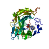
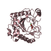
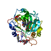
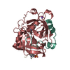
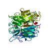
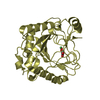
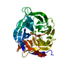
| Title | Crystal structure of a putative, de novo designed unnatural amino acid dependent metalloprotein, northeast structural genomics consortium target OR61 | ||||||
 Components Components | designed unnatural amino acid dependent metalloprotein | ||||||
 Keywords Keywords | Structural Genomics / Unknown Function / PSI-Biology / Northeast Structural Genomics Consortium / NESG / a beta-propeller / novel metalloenzyme | ||||||
| Function / homology |  Function and homology information Function and homology informationganglioside catabolic process / oligosaccharide catabolic process / exo-alpha-sialidase / exo-alpha-sialidase activity / extracellular region / membrane / cytoplasm Similarity search - Function | ||||||
| Biological species |  Micromonospora viridifaciens (bacteria) Micromonospora viridifaciens (bacteria) | ||||||
| Method |  X-RAY DIFFRACTION / SYNCHROTRON / MOLECULAR REPLACEMENT / Resolution: 1.4 Å X-RAY DIFFRACTION / SYNCHROTRON / MOLECULAR REPLACEMENT / Resolution: 1.4 Å | ||||||
 Authors Authors | Forouhar, F. / Lew, S. / Seetharaman, J. / Mills, J.H. / Khare, S.D. / Everett, J.K. / Baker, D. / Montelione, G.T. / Hunt, J.F. / Tong, L. / Northeast Structural Genomics Consortium (NESG) | ||||||
 Citation Citation | Journal: J.Am.Chem.Soc. / Year: 2013 Title: Computational design of an unnatural amino Acid dependent metalloprotein with atomic level accuracy. Authors: Mills, J.H. / Khare, S.D. / Bolduc, J.M. / Forouhar, F. / Mulligan, V.K. / Lew, S. / Seetharaman, J. / Tong, L. / Stoddard, B.L. / Baker, D. | ||||||
| History |
|
- Structure visualization
| Structure viewer | Molecule: MolmilJmol/JSmol |
|---|
- Downloads & links
-Download
| PDBx/mmCIF format | 4j9t.cif.gz | 169.5 KB | Display | PDBx/mmCIF format |
|---|---|---|---|---|
| PDB format | pdb4j9t.ent.gz | 130.1 KB | Display | PDB format |
| PDBx/mmJSON format | 4j9t.json.gz | Tree view | PDBx/mmJSON format | |
| Others |  Other downloads Other downloads |
-Validation report
| Arichive directory | https://data.pdbj.org/pub/pdb/validation_reports/j9/4j9tftp://data.pdbj.org/pub/pdb/validation_reports/j9/4j9t | HTTPS FTP |
|---|
-Related structure data
| Related structure data | 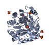 4iwwC 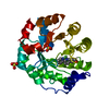 4ix0C 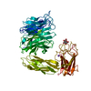 1euuS S: Starting model for refinement C: citing same article ( |
|---|---|
| Similar structure data | |
| Other databases |


-Links
 PDBj
PDBj


- Assembly
| Deposited unit | 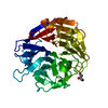
| ||||||||
|---|---|---|---|---|---|---|---|---|---|
| 1 |
| ||||||||
| Unit cell |
|
-Components
| #1: Protein | Mass: 38549.270 Da / Num. of mol.: 1 Mutation: R68S, I69H, D92A, D131H, F155W, A156E, T226A, F234I, D259S, E260H, R276A, N311D Source method: isolated from a genetically manipulated source Source: (gene. exp.) Micromonospora viridifaciens (bacteria)Gene: nedA / Plasmid: pET29b / Production host: |
|---|---|
| #2: Chemical | ChemComp-GOL /   Mass: 92.094 Da / Num. of mol.: 1 / Source method: obtained synthetically / Formula: C3H8O3 Mass: 92.094 Da / Num. of mol.: 1 / Source method: obtained synthetically / Formula: C3H8O3 |
| #3: Chemical | ChemComp-ARS /   Mass: 74.922 Da / Num. of mol.: 1 / Source method: obtained synthetically / Formula: As Mass: 74.922 Da / Num. of mol.: 1 / Source method: obtained synthetically / Formula: As |
| #4: Water | ChemComp-HOH /  Mass: 18.015 Da / Num. of mol.: 527 / Source method: isolated from a natural source / Formula: H2O Mass: 18.015 Da / Num. of mol.: 527 / Source method: isolated from a natural source / Formula: H2O |
| Compound details | residue 92, which is modeled as Ala in the current structure, is modified by unnatural amino acid ...residue 92, which is modeled as Ala in the current structure, is modified by unnatural amino acid biprimidine Ala. |
-Experimental details
-Experiment
| Experiment | Method: X-RAY DIFFRACTION / Number of used crystals: 1 |
|---|
- Sample preparation
| Crystal | Density Matthews: 2 Å3/Da / Density % sol: 38.45 % |
|---|---|
| Crystal grow | Temperature: 277 K / Method: microbatch under oil / pH: 6.5 Details: protein buffer: 10 mM Tris HCl (pH 8). Precipitation cocktail: 100 MM NA CACODYLATE (PH 6.5), 5 mM FeCl2, 15% PEG 3350 Microbatch under oil, temperature 277K |
-Data collection
| Diffraction | Mean temperature: 100 K |
|---|---|
| Diffraction source | Source: SYNCHROTRON / Site: NSLS  / Beamline: X4C / Wavelength: 0.97012 Å / Beamline: X4C / Wavelength: 0.97012 Å |
| Detector | Type: MAR CCD 165 mm / Detector: CCD / Date: Oct 12, 2012 / Details: mirrors |
| Radiation | Monochromator: Si 111 CHANNEL / Protocol: SINGLE WAVELENGTH / Monochromatic (M) / Laue (L): M / Scattering type: x-ray |
| Radiation wavelength | Wavelength: 0.97012 Å / Relative weight: 1 |
| Reflection | Resolution: 1.4→31.021 Å / Num. obs: 61532 / % possible obs: 99.9 % / Observed criterion σ(F): 0 / Observed criterion σ(I): 0 / Redundancy: 6.1 % / Biso Wilson estimate: 11.2 Å2 / Rmerge(I) obs: 0.059 / Rsym value: 0.041 / Net I/σ(I): 29.2 |
| Reflection shell | Resolution: 1.4→1.45 Å / Redundancy: 4.9 % / Rmerge(I) obs: 0.42 / Mean I/σ(I) obs: 4.9 / Num. unique all: 6060 / Rsym value: 0.405 / % possible all: 99.7 |
- Processing
| Software |
| |||||||||||||||||||||||||||||||||||||||||||||||||||||||||||||||||||||||||||||||||||||||||||||||||||||||||||||||||||||||||||||||||||||||||||||||||||||||||||||||||||||||||||||||||||||||||||||||||||||||||||||||||||||||||
|---|---|---|---|---|---|---|---|---|---|---|---|---|---|---|---|---|---|---|---|---|---|---|---|---|---|---|---|---|---|---|---|---|---|---|---|---|---|---|---|---|---|---|---|---|---|---|---|---|---|---|---|---|---|---|---|---|---|---|---|---|---|---|---|---|---|---|---|---|---|---|---|---|---|---|---|---|---|---|---|---|---|---|---|---|---|---|---|---|---|---|---|---|---|---|---|---|---|---|---|---|---|---|---|---|---|---|---|---|---|---|---|---|---|---|---|---|---|---|---|---|---|---|---|---|---|---|---|---|---|---|---|---|---|---|---|---|---|---|---|---|---|---|---|---|---|---|---|---|---|---|---|---|---|---|---|---|---|---|---|---|---|---|---|---|---|---|---|---|---|---|---|---|---|---|---|---|---|---|---|---|---|---|---|---|---|---|---|---|---|---|---|---|---|---|---|---|---|---|---|---|---|---|---|---|---|---|---|---|---|---|---|---|---|---|---|---|---|---|
| Refinement | Method to determine structure: MOLECULAR REPLACEMENT Starting model: PDB ENTRY 1EUU Resolution: 1.4→31.021 Å / SU ML: 0.18 / Cross valid method: THROUGHOUT / σ(F): 1.34 / σ(I): 1.3 / Phase error: 13.94 / Stereochemistry target values: ML Details: the electron density near Cys-351 was modeled as As ion, however, Fe2+ could be another possibility
| |||||||||||||||||||||||||||||||||||||||||||||||||||||||||||||||||||||||||||||||||||||||||||||||||||||||||||||||||||||||||||||||||||||||||||||||||||||||||||||||||||||||||||||||||||||||||||||||||||||||||||||||||||||||||
| Solvent computation | Shrinkage radii: 0.83 Å / VDW probe radii: 1.1 Å / Solvent model: FLAT BULK SOLVENT MODEL / Bsol: 40.338 Å2 / ksol: 0.396 e/Å3 | |||||||||||||||||||||||||||||||||||||||||||||||||||||||||||||||||||||||||||||||||||||||||||||||||||||||||||||||||||||||||||||||||||||||||||||||||||||||||||||||||||||||||||||||||||||||||||||||||||||||||||||||||||||||||
| Displacement parameters |
| |||||||||||||||||||||||||||||||||||||||||||||||||||||||||||||||||||||||||||||||||||||||||||||||||||||||||||||||||||||||||||||||||||||||||||||||||||||||||||||||||||||||||||||||||||||||||||||||||||||||||||||||||||||||||
| Refine analyze |
| |||||||||||||||||||||||||||||||||||||||||||||||||||||||||||||||||||||||||||||||||||||||||||||||||||||||||||||||||||||||||||||||||||||||||||||||||||||||||||||||||||||||||||||||||||||||||||||||||||||||||||||||||||||||||
| Refinement step | Cycle: LAST / Resolution: 1.4→31.021 Å
| |||||||||||||||||||||||||||||||||||||||||||||||||||||||||||||||||||||||||||||||||||||||||||||||||||||||||||||||||||||||||||||||||||||||||||||||||||||||||||||||||||||||||||||||||||||||||||||||||||||||||||||||||||||||||
| Refine LS restraints |
| |||||||||||||||||||||||||||||||||||||||||||||||||||||||||||||||||||||||||||||||||||||||||||||||||||||||||||||||||||||||||||||||||||||||||||||||||||||||||||||||||||||||||||||||||||||||||||||||||||||||||||||||||||||||||
| LS refinement shell |
|
