 Movie
Movie Controller

[English] 日本語
 Yorodumi
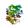
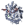
- PDB-3vdh: Crystal structure of PbGH5A, a glycoside hydrolase family 5 enzym... -
+ Open data

- Basic information
| Entry | Database: PDB / ID: 3vdh | |||||||||
|---|---|---|---|---|---|---|---|---|---|---|
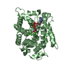
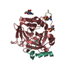
| Title | Crystal structure of PbGH5A, a glycoside hydrolase family 5 enzyme from Prevotella bryantii B14 | |||||||||
 Components Components | B-1,4-endoglucanase | |||||||||
 Keywords Keywords | HYDROLASE / ENDO-BETA-GLUCANASE/ENDO-XYLOGLUCANASE / GLYCOSYL HYDROLASE FAMILY 5 / MIXED ALPHA-BETA / TIM BARREL / STRUCTURAL GENOMICS / PSI-BIOLOGY / PROTEIN STRUCTURE INITIATIVE / MIDWEST CENTER FOR STRUCTURAL GENOMICS / MCSG | |||||||||
| Function / homology |  Function and homology information Function and homology informationsubstituted mannan metabolic process / mannan endo-1,4-beta-mannosidase activity / polysaccharide catabolic process / metal ion binding Similarity search - Function | |||||||||
| Biological species |  Prevotella bryantii (bacteria) Prevotella bryantii (bacteria) | |||||||||
| Method |  X-RAY DIFFRACTION / MOLECULAR REPLACEMENT / Resolution: 1.62 Å X-RAY DIFFRACTION / MOLECULAR REPLACEMENT / Resolution: 1.62 Å | |||||||||
 Authors Authors | Stogios, P.J. / Evdokimova, E. / Egorova, O. / Yim, V. / Joachimiak, A. / Edwards, A.M. / Savchenko, A. / Midwest Center for Structural Genomics (MCSG) | |||||||||
 Citation Citation | Journal: J.Biol.Chem. / Year: 2016 Title: Structure-Function Analysis of a Mixed-linkage beta-Glucanase/Xyloglucanase from the Key Ruminal Bacteroidetes Prevotella bryantii B14. Authors: McGregor, N. / Morar, M. / Fenger, T.H. / Stogios, P. / Lenfant, N. / Yin, V. / Xu, X. / Evdokimova, E. / Cui, H. / Henrissat, B. / Savchenko, A. / Brumer, H. | |||||||||
| History |
|
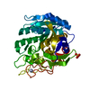
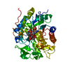
- Structure visualization
| Structure viewer | Molecule: MolmilJmol/JSmol |
|---|
- Downloads & links
-Download
| PDBx/mmCIF format | 3vdh.cif.gz | 325.4 KB | Display | PDBx/mmCIF format |
|---|---|---|---|---|
| PDB format | pdb3vdh.ent.gz | 268.1 KB | Display | PDB format |
| PDBx/mmJSON format | 3vdh.json.gz | Tree view | PDBx/mmJSON format | |
| Others |  Other downloads Other downloads |
-Validation report
| Arichive directory | https://data.pdbj.org/pub/pdb/validation_reports/vd/3vdhftp://data.pdbj.org/pub/pdb/validation_reports/vd/3vdh | HTTPS FTP |
|---|
-Related structure data
| Related structure data | 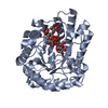 5d9mC 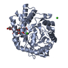 5d9nC 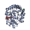 5d9oC  5d9pC  2jeqS C: citing same article ( S: Starting model for refinement |
|---|---|
| Similar structure data | |
| Other databases |


-Links
 PDBj
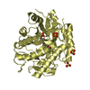
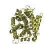
PDBj- Assembly
| Deposited unit | 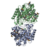
| ||||||||
|---|---|---|---|---|---|---|---|---|---|
| 1 | 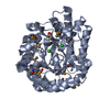
| ||||||||
| 2 | 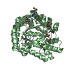
| ||||||||
| Unit cell |
|
-Components
| #1: Protein | Mass: 40044.039 Da / Num. of mol.: 2 Source method: isolated from a genetically manipulated source Source: (gene. exp.) Prevotella bryantii (bacteria) / Plasmid: PET101-11079 / Production host: #2: Chemical |   Mass: 35.453 Da / Num. of mol.: 3 / Source method: obtained synthetically / Formula: Cl Mass: 35.453 Da / Num. of mol.: 3 / Source method: obtained synthetically / Formula: Cl#3: Water | ChemComp-HOH / |  Mass: 18.015 Da / Num. of mol.: 912 / Source method: isolated from a natural source / Formula: H2O Mass: 18.015 Da / Num. of mol.: 912 / Source method: isolated from a natural source / Formula: H2OHas protein modification | Y | |
|---|
-Experimental details
-Experiment
| Experiment | Method: X-RAY DIFFRACTION / Number of used crystals: 1 |
|---|
- Sample preparation
| Crystal | Density Matthews: 2.17 Å3/Da / Density % sol: 43.34 % |
|---|---|
| Crystal grow | Temperature: 295 K / Method: vapor diffusion, hanging drop / pH: 6.5 Details: 0.2 M CALCIUM ACETATE HYDRATE, 0.1M SODIUM CACODYLATE PH 6.5, 18% PEG 8K, VAPOR DIFFUSION, HANGING DROP, temperature 295K |
-Data collection
| Diffraction | Mean temperature: 100 K |
|---|---|
| Diffraction source | Source: ROTATING ANODE / Type: RIGAKU MICROMAX-007 / Wavelength: 1.54178 Å |
| Detector | Type: RIGAKU RAXIS IV / Detector: IMAGE PLATE / Date: Oct 22, 2009 / Details: mirrors |
| Radiation | Monochromator: graphite / Protocol: SINGLE WAVELENGTH / Monochromatic (M) / Laue (L): M / Scattering type: x-ray |
| Radiation wavelength | Wavelength: 1.54178 Å / Relative weight: 1 |
| Reflection | Resolution: 1.55→50 Å / Num. obs: 83351 / % possible obs: 84 % / Observed criterion σ(F): 0 / Observed criterion σ(I): -2 / Redundancy: 3.4 % / Rsym value: 0.039 / Net I/σ(I): 38.95 |
| Reflection shell | Resolution: 1.6→1.63 Å / Redundancy: 4 % / Mean I/σ(I) obs: 4.67 / Rsym value: 0.443 / % possible all: 61.6 |
- Processing
| Software |
| |||||||||||||||||||||||||||||||||||||||||||||||||||||||||||||||||||||||||||||||||||||||||||||||||||||||||||||||||||||||||||||||||||||||||||||||||||||||||||||||||||||||||||||||
|---|---|---|---|---|---|---|---|---|---|---|---|---|---|---|---|---|---|---|---|---|---|---|---|---|---|---|---|---|---|---|---|---|---|---|---|---|---|---|---|---|---|---|---|---|---|---|---|---|---|---|---|---|---|---|---|---|---|---|---|---|---|---|---|---|---|---|---|---|---|---|---|---|---|---|---|---|---|---|---|---|---|---|---|---|---|---|---|---|---|---|---|---|---|---|---|---|---|---|---|---|---|---|---|---|---|---|---|---|---|---|---|---|---|---|---|---|---|---|---|---|---|---|---|---|---|---|---|---|---|---|---|---|---|---|---|---|---|---|---|---|---|---|---|---|---|---|---|---|---|---|---|---|---|---|---|---|---|---|---|---|---|---|---|---|---|---|---|---|---|---|---|---|---|---|---|---|
| Refinement | Method to determine structure: MOLECULAR REPLACEMENT Starting model: pdb entry 2JEQ Resolution: 1.62→26.354 Å / SU ML: 0.25 / Cross valid method: THROUGHOUT / σ(F): 1.34 / Phase error: 21.22 / Stereochemistry target values: MLHL
| |||||||||||||||||||||||||||||||||||||||||||||||||||||||||||||||||||||||||||||||||||||||||||||||||||||||||||||||||||||||||||||||||||||||||||||||||||||||||||||||||||||||||||||||
| Solvent computation | Shrinkage radii: 0.9 Å / VDW probe radii: 1.11 Å / Solvent model: FLAT BULK SOLVENT MODEL / Bsol: 55.549 Å2 / ksol: 0.354 e/Å3 | |||||||||||||||||||||||||||||||||||||||||||||||||||||||||||||||||||||||||||||||||||||||||||||||||||||||||||||||||||||||||||||||||||||||||||||||||||||||||||||||||||||||||||||||
| Displacement parameters |
| |||||||||||||||||||||||||||||||||||||||||||||||||||||||||||||||||||||||||||||||||||||||||||||||||||||||||||||||||||||||||||||||||||||||||||||||||||||||||||||||||||||||||||||||
| Refinement step | Cycle: LAST / Resolution: 1.62→26.354 Å
| |||||||||||||||||||||||||||||||||||||||||||||||||||||||||||||||||||||||||||||||||||||||||||||||||||||||||||||||||||||||||||||||||||||||||||||||||||||||||||||||||||||||||||||||
| Refine LS restraints |
| |||||||||||||||||||||||||||||||||||||||||||||||||||||||||||||||||||||||||||||||||||||||||||||||||||||||||||||||||||||||||||||||||||||||||||||||||||||||||||||||||||||||||||||||
| LS refinement shell |
| |||||||||||||||||||||||||||||||||||||||||||||||||||||||||||||||||||||||||||||||||||||||||||||||||||||||||||||||||||||||||||||||||||||||||||||||||||||||||||||||||||||||||||||||
| Refinement TLS params. | Method: refined / Refine-ID: X-RAY DIFFRACTION
| |||||||||||||||||||||||||||||||||||||||||||||||||||||||||||||||||||||||||||||||||||||||||||||||||||||||||||||||||||||||||||||||||||||||||||||||||||||||||||||||||||||||||||||||
| Refinement TLS group |
|
