 Movie
Movie Controller

[English] 日本語
 Yorodumi
- PDB-5d9o: Crystal structure of PbGH5A, a glycoside hydrolase family 5 enzym... -
+ Open data

- Basic information
| Entry | Database: PDB / ID: 5d9o | |||||||||
|---|---|---|---|---|---|---|---|---|---|---|
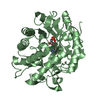
| Title | Crystal structure of PbGH5A, a glycoside hydrolase family 5 enzyme from Prevotella bryantii B14, E280A mutant in complex with cellotetraose | |||||||||
 Components Components | B-1,4-endoglucanase | |||||||||
 Keywords Keywords | HYDROLASE / endo-beta-glucanase/endo-xyloglucanase / GLYCOSYL HYDROLASE FAMILY 5 / MIXED ALPHA-BETA / TIM BARREL | |||||||||
| Function / homology |  Function and homology information Function and homology informationsubstituted mannan metabolic process / mannan endo-1,4-beta-mannosidase activity / polysaccharide catabolic process / metal ion binding Similarity search - Function | |||||||||
| Biological species |  Prevotella bryantii (bacteria) Prevotella bryantii (bacteria) | |||||||||
| Method |  X-RAY DIFFRACTION / MOLECULAR REPLACEMENT / Resolution: 1.55 Å X-RAY DIFFRACTION / MOLECULAR REPLACEMENT / Resolution: 1.55 Å | |||||||||
 Authors Authors | Morar, M. / Stogios, P.J. / Xu, X. / Cui, H. / Di Leo, R. / Yim, V. / Savchenko, A. | |||||||||
 Citation Citation | Journal: J.Biol.Chem. / Year: 2016 Title: Structure-Function Analysis of a Mixed-linkage beta-Glucanase/Xyloglucanase from the Key Ruminal Bacteroidetes Prevotella bryantii B14. Authors: McGregor, N. / Morar, M. / Fenger, T.H. / Stogios, P. / Lenfant, N. / Yin, V. / Xu, X. / Evdokimova, E. / Cui, H. / Henrissat, B. / Savchenko, A. / Brumer, H. | |||||||||
| History |
|
- Structure visualization
| Structure viewer | Molecule: MolmilJmol/JSmol |
|---|
- Downloads & links
-Download
| PDBx/mmCIF format | 5d9o.cif.gz | 322.4 KB | Display | PDBx/mmCIF format |
|---|---|---|---|---|
| PDB format | pdb5d9o.ent.gz | 259.8 KB | Display | PDB format |
| PDBx/mmJSON format | 5d9o.json.gz | Tree view | PDBx/mmJSON format | |
| Others |  Other downloads Other downloads |
-Validation report
| Arichive directory | https://data.pdbj.org/pub/pdb/validation_reports/d9/5d9oftp://data.pdbj.org/pub/pdb/validation_reports/d9/5d9o | HTTPS FTP |
|---|
-Related structure data
| Related structure data | 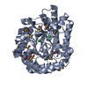 3vdhSC 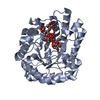 5d9mC 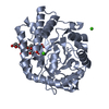 5d9nC 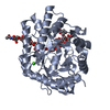 5d9pC S: Starting model for refinement C: citing same article ( |
|---|---|
| Similar structure data |

-Links
 PDBj
PDBj- Assembly
| Deposited unit | 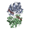
| ||||||||
|---|---|---|---|---|---|---|---|---|---|
| 1 | 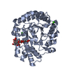
| ||||||||
| 2 | 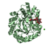
| ||||||||
| Unit cell |
|
-Components
| #1: Protein | Mass: 39329.473 Da / Num. of mol.: 2 / Mutation: E280A Source method: isolated from a genetically manipulated source Source: (gene. exp.) Prevotella bryantii (bacteria) / Plasmid: p15TV-LIC / Production host: #2: Polysaccharide |   Source method: isolated from a genetically manipulated source Details: oligosaccharide / References: beta-cellotetraose #3: Chemical |   Mass: 40.078 Da / Num. of mol.: 3 / Source method: obtained synthetically / Formula: Ca Mass: 40.078 Da / Num. of mol.: 3 / Source method: obtained synthetically / Formula: Ca#4: Water | ChemComp-HOH / |  Mass: 18.015 Da / Num. of mol.: 987 / Source method: isolated from a natural source / Formula: H2O Mass: 18.015 Da / Num. of mol.: 987 / Source method: isolated from a natural source / Formula: H2O |
|---|
-Experimental details
-Experiment
| Experiment | Method: X-RAY DIFFRACTION |
|---|
- Sample preparation
| Crystal | Density Matthews: 1.95 Å3/Da / Density % sol: 36.97 % |
|---|---|
| Crystal grow | Temperature: 298 K / Method: vapor diffusion, hanging drop Details: 1.8 microL of protein solution at 28 mg/mL mixed with 1.8 microL of reservoir solution (0.1 M sodium cacodylate pH 6.3 to 7.1, 0.2 M calcium acetate, 25% PEG8K), then soaking crystals in ...Details: 1.8 microL of protein solution at 28 mg/mL mixed with 1.8 microL of reservoir solution (0.1 M sodium cacodylate pH 6.3 to 7.1, 0.2 M calcium acetate, 25% PEG8K), then soaking crystals in reservoir solution supplemented with 10 mM cellotetraose for 2 h. Cryoprotectant = paratone-N oil. |
-Data collection
| Diffraction | Mean temperature: 100 K |
|---|---|
| Diffraction source | Source: ROTATING ANODE / Type: RIGAKU MICROMAX-007 HF / Wavelength: 1.5418 Å |
| Detector | Type: RIGAKU SATURN A200 / Detector: CCD / Date: Oct 22, 2014 |
| Radiation | Protocol: SINGLE WAVELENGTH / Monochromatic (M) / Laue (L): M / Scattering type: x-ray |
| Radiation wavelength | Wavelength: 1.5418 Å / Relative weight: 1 |
| Reflection | Resolution: 1.55→19.47 Å / Num. obs: 85184 / % possible obs: 97.1 % / Redundancy: 4 % / Rsym value: 0.082 / Net I/σ(I): 14 |
| Reflection shell | Resolution: 1.55→1.63 Å / Redundancy: 4 % / Rmerge(I) obs: 0.535 / Mean I/σ(I) obs: 2.9 / % possible all: 94.1 |
- Processing
| Software |
| |||||||||||||||||||||||||||||||||||||||||||||||||||||||||||||||||||||||||||||||||||||||||||||||||||||||||||||||||||||||||||||||||||||||||||||||||||||||||||||||||||||||||||||||||||||||||||||||||||||||||||||||||||||||||
|---|---|---|---|---|---|---|---|---|---|---|---|---|---|---|---|---|---|---|---|---|---|---|---|---|---|---|---|---|---|---|---|---|---|---|---|---|---|---|---|---|---|---|---|---|---|---|---|---|---|---|---|---|---|---|---|---|---|---|---|---|---|---|---|---|---|---|---|---|---|---|---|---|---|---|---|---|---|---|---|---|---|---|---|---|---|---|---|---|---|---|---|---|---|---|---|---|---|---|---|---|---|---|---|---|---|---|---|---|---|---|---|---|---|---|---|---|---|---|---|---|---|---|---|---|---|---|---|---|---|---|---|---|---|---|---|---|---|---|---|---|---|---|---|---|---|---|---|---|---|---|---|---|---|---|---|---|---|---|---|---|---|---|---|---|---|---|---|---|---|---|---|---|---|---|---|---|---|---|---|---|---|---|---|---|---|---|---|---|---|---|---|---|---|---|---|---|---|---|---|---|---|---|---|---|---|---|---|---|---|---|---|---|---|---|---|---|---|---|
| Refinement | Method to determine structure: MOLECULAR REPLACEMENT Starting model: 3VDH Resolution: 1.55→19.456 Å / SU ML: 0.14 / Cross valid method: FREE R-VALUE / σ(F): 1.34 / Phase error: 16.42 / Stereochemistry target values: ML
| |||||||||||||||||||||||||||||||||||||||||||||||||||||||||||||||||||||||||||||||||||||||||||||||||||||||||||||||||||||||||||||||||||||||||||||||||||||||||||||||||||||||||||||||||||||||||||||||||||||||||||||||||||||||||
| Solvent computation | Shrinkage radii: 0.9 Å / VDW probe radii: 1.11 Å / Solvent model: FLAT BULK SOLVENT MODEL | |||||||||||||||||||||||||||||||||||||||||||||||||||||||||||||||||||||||||||||||||||||||||||||||||||||||||||||||||||||||||||||||||||||||||||||||||||||||||||||||||||||||||||||||||||||||||||||||||||||||||||||||||||||||||
| Refinement step | Cycle: LAST / Resolution: 1.55→19.456 Å
| |||||||||||||||||||||||||||||||||||||||||||||||||||||||||||||||||||||||||||||||||||||||||||||||||||||||||||||||||||||||||||||||||||||||||||||||||||||||||||||||||||||||||||||||||||||||||||||||||||||||||||||||||||||||||
| Refine LS restraints |
| |||||||||||||||||||||||||||||||||||||||||||||||||||||||||||||||||||||||||||||||||||||||||||||||||||||||||||||||||||||||||||||||||||||||||||||||||||||||||||||||||||||||||||||||||||||||||||||||||||||||||||||||||||||||||
| LS refinement shell |
| |||||||||||||||||||||||||||||||||||||||||||||||||||||||||||||||||||||||||||||||||||||||||||||||||||||||||||||||||||||||||||||||||||||||||||||||||||||||||||||||||||||||||||||||||||||||||||||||||||||||||||||||||||||||||
| Refinement TLS params. | Method: refined / Origin x: -3.1645 Å / Origin y: -17.5881 Å / Origin z: -43.8681 Å
| |||||||||||||||||||||||||||||||||||||||||||||||||||||||||||||||||||||||||||||||||||||||||||||||||||||||||||||||||||||||||||||||||||||||||||||||||||||||||||||||||||||||||||||||||||||||||||||||||||||||||||||||||||||||||
| Refinement TLS group | Selection details: all |
