 Movie
Movie Controller

[English] 日本語
 Yorodumi
- PDB-4psu: Crystal structure of alpha/beta hydrolase from Rhodopseudomonas p... -
+ Open data

- Basic information
| Entry | Database: PDB / ID: 4psu | ||||||
|---|---|---|---|---|---|---|---|
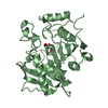
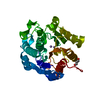
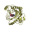
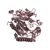
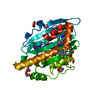
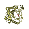
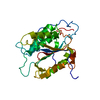
| Title | Crystal structure of alpha/beta hydrolase from Rhodopseudomonas palustris CGA009 | ||||||
 Components Components | Alpha/beta hydrolase | ||||||
 Keywords Keywords | HYDROLASE / alpha/beta hydrolase / esterase | ||||||
| Function / homology |  Function and homology information Function and homology information | ||||||
| Biological species |  Rhodopseudomonas palustris (phototrophic) Rhodopseudomonas palustris (phototrophic) | ||||||
| Method |  X-RAY DIFFRACTION / SYNCHROTRON / SAD / Resolution: 2.2 Å X-RAY DIFFRACTION / SYNCHROTRON / SAD / Resolution: 2.2 Å | ||||||
 Authors Authors | Nocek, B. / Hajighasemi, M. / Xu, X. / Cui, H. / Savchenko, A. / Yakunin, A. | ||||||
 Citation Citation | Journal: TO BE PUBLISHED Title: Crystal structure of alpha/beta hydrolase from Rhodopseudomonas palustris CGA009 Authors: Nocek, B. / Hajighasemi, M. / Xu, X. / Cui, H. / Savchenko, A. / Yakunin, A. | ||||||
| History |
|
- Structure visualization
| Structure viewer | Molecule: MolmilJmol/JSmol |
|---|
- Downloads & links
-Download
| PDBx/mmCIF format | 4psu.cif.gz | 123.7 KB | Display | PDBx/mmCIF format |
|---|---|---|---|---|
| PDB format | pdb4psu.ent.gz | 95.9 KB | Display | PDB format |
| PDBx/mmJSON format | 4psu.json.gz | Tree view | PDBx/mmJSON format | |
| Others |  Other downloads Other downloads |
-Validation report
| Arichive directory | https://data.pdbj.org/pub/pdb/validation_reports/ps/4psuftp://data.pdbj.org/pub/pdb/validation_reports/ps/4psu | HTTPS FTP |
|---|
-Related structure data
| Similar structure data |
|---|


-Links
 PDBj
PDBj

- Assembly
| Deposited unit | 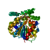
| ||||||||
|---|---|---|---|---|---|---|---|---|---|
| 1 |
| ||||||||
| Unit cell |
|
-Components
| #1: Protein | Mass: 32181.133 Da / Num. of mol.: 1 Source method: isolated from a genetically manipulated source Source: (gene. exp.) Rhodopseudomonas palustris (phototrophic)Strain: ATCC BAA-98 / CGA009 / Gene: RPA1511 / Production host: |
|---|---|
| #2: Chemical | ChemComp-12P /   Mass: 546.646 Da / Num. of mol.: 1 / Source method: obtained synthetically / Formula: C24H50O13 / Comment: precipitant*YM Mass: 546.646 Da / Num. of mol.: 1 / Source method: obtained synthetically / Formula: C24H50O13 / Comment: precipitant*YM |
| #3: Water | ChemComp-HOH /  Mass: 18.015 Da / Num. of mol.: 69 / Source method: isolated from a natural source / Formula: H2O Mass: 18.015 Da / Num. of mol.: 69 / Source method: isolated from a natural source / Formula: H2O |
| Has protein modification | Y |
-Experimental details
-Experiment
| Experiment | Method: X-RAY DIFFRACTION / Number of used crystals: 1 |
|---|
- Sample preparation
| Crystal | Density Matthews: 2.55 Å3/Da / Density % sol: 51.69 % |
|---|---|
| Crystal grow | Temperature: 289 K / Method: vapor diffusion, hanging drop / pH: 7 Details: 0.2 M potassium thiocyanate, 20% PEG3350, 4% Jeffmine M-600, 1/300 trypsin, pH 7.0, VAPOR DIFFUSION, HANGING DROP, temperature 289K |
-Data collection
| Diffraction | Mean temperature: 100 K |
|---|---|
| Diffraction source | Source: SYNCHROTRON / Site: APS  / Beamline: 19-ID / Wavelength: 0.9794 Å / Beamline: 19-ID / Wavelength: 0.9794 Å |
| Detector | Type: ADSC QUANTUM 315r / Detector: CCD / Date: Oct 20, 2013 / Details: mirrors |
| Radiation | Monochromator: double crystal Si(111) / Protocol: SINGLE WAVELENGTH / Monochromatic (M) / Laue (L): M / Scattering type: x-ray |
| Radiation wavelength | Wavelength: 0.9794 Å / Relative weight: 1 |
| Reflection | Resolution: 2.2→32 Å / Num. all: 16900 / Num. obs: 16885 / % possible obs: 99.3 % / Observed criterion σ(F): 2 / Observed criterion σ(I): 2 / Redundancy: 3.8 % / Rmerge(I) obs: 0.077 / Net I/σ(I): 15 |
| Reflection shell | Resolution: 2.2→2.24 Å / % possible all: 98.1 |
- Processing
| Software |
| |||||||||||||||||||||||||||||||||||||||||||||||||||||||||||||||||||||||||||||||||||||||||||||||||||||||||||||||||||||||||||||
|---|---|---|---|---|---|---|---|---|---|---|---|---|---|---|---|---|---|---|---|---|---|---|---|---|---|---|---|---|---|---|---|---|---|---|---|---|---|---|---|---|---|---|---|---|---|---|---|---|---|---|---|---|---|---|---|---|---|---|---|---|---|---|---|---|---|---|---|---|---|---|---|---|---|---|---|---|---|---|---|---|---|---|---|---|---|---|---|---|---|---|---|---|---|---|---|---|---|---|---|---|---|---|---|---|---|---|---|---|---|---|---|---|---|---|---|---|---|---|---|---|---|---|---|---|---|---|
| Refinement | Method to determine structure: SAD / Resolution: 2.2→31.961 Å / SU ML: 0.25 / σ(F): 2 / Phase error: 23.77 / Stereochemistry target values: ML
| |||||||||||||||||||||||||||||||||||||||||||||||||||||||||||||||||||||||||||||||||||||||||||||||||||||||||||||||||||||||||||||
| Solvent computation | Shrinkage radii: 0.9 Å / VDW probe radii: 1.11 Å / Solvent model: FLAT BULK SOLVENT MODEL | |||||||||||||||||||||||||||||||||||||||||||||||||||||||||||||||||||||||||||||||||||||||||||||||||||||||||||||||||||||||||||||
| Refinement step | Cycle: LAST / Resolution: 2.2→31.961 Å
| |||||||||||||||||||||||||||||||||||||||||||||||||||||||||||||||||||||||||||||||||||||||||||||||||||||||||||||||||||||||||||||
| Refine LS restraints |
| |||||||||||||||||||||||||||||||||||||||||||||||||||||||||||||||||||||||||||||||||||||||||||||||||||||||||||||||||||||||||||||
| LS refinement shell |
| |||||||||||||||||||||||||||||||||||||||||||||||||||||||||||||||||||||||||||||||||||||||||||||||||||||||||||||||||||||||||||||
| Refinement TLS params. | Method: refined / Refine-ID: X-RAY DIFFRACTION
| |||||||||||||||||||||||||||||||||||||||||||||||||||||||||||||||||||||||||||||||||||||||||||||||||||||||||||||||||||||||||||||
| Refinement TLS group |
|
