 Movie
Movie Controller

[English] 日本語
 Yorodumi
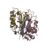
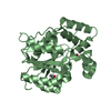
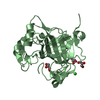
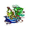
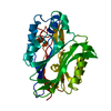
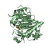
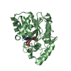
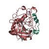
- PDB-3ep6: Human AdoMetDC D174N mutant complexed with S-Adenosylmethionine m... -
+ Open data

- Basic information
| Entry | Database: PDB / ID: 3ep6 | |||||||||
|---|---|---|---|---|---|---|---|---|---|---|
| Title | Human AdoMetDC D174N mutant complexed with S-Adenosylmethionine methyl ester and no putrescine bound | |||||||||
 Components Components |
| |||||||||
 Keywords Keywords | LYASE / AdoMetDC with mutation in putrescine binding site / Decarboxylase / Pyruvate / S-adenosyl-L-methionine / Spermidine biosynthesis / Zymogen | |||||||||
| Function / homology |  Function and homology information Function and homology informationspermine biosynthetic process / adenosylmethionine decarboxylase / adenosylmethionine decarboxylase activity / Metabolism of polyamines / polyamine metabolic process / putrescine binding / spermidine biosynthetic process / identical protein binding / cytosol Similarity search - Function | |||||||||
| Biological species |  Homo sapiens (human) Homo sapiens (human) | |||||||||
| Method |  X-RAY DIFFRACTION / SYNCHROTRON / MOLECULAR REPLACEMENT / Resolution: 1.7 Å X-RAY DIFFRACTION / SYNCHROTRON / MOLECULAR REPLACEMENT / Resolution: 1.7 Å | |||||||||
 Authors Authors | Bale, S. / Lopez, M.M. / Makhatadze, G.I. / Fang, Q. / Pegg, A.E. / Ealick, S.E. | |||||||||
 Citation Citation | Journal: Biochemistry / Year: 2008 Title: Structural Basis for Putrescine Activation of Human S-Adenosylmethionine Decarboxylase. Authors: Bale, S. / Lopez, M.M. / Makhatadze, G.I. / Fang, Q. / Pegg, A.E. / Ealick, S.E. | |||||||||
| History |
|
- Structure visualization
| Structure viewer | Molecule: MolmilJmol/JSmol |
|---|
- Downloads & links
-Download
| PDBx/mmCIF format | 3ep6.cif.gz | 81.2 KB | Display | PDBx/mmCIF format |
|---|---|---|---|---|
| PDB format | pdb3ep6.ent.gz | 57.6 KB | Display | PDB format |
| PDBx/mmJSON format | 3ep6.json.gz | Tree view | PDBx/mmJSON format | |
| Others |  Other downloads Other downloads |
-Validation report
| Arichive directory | https://data.pdbj.org/pub/pdb/validation_reports/ep/3ep6ftp://data.pdbj.org/pub/pdb/validation_reports/ep/3ep6 | HTTPS FTP |
|---|
-Related structure data
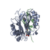
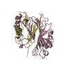
| Related structure data |  3ep3C  3ep4C 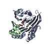 3ep5C  3ep7C  3ep8C 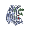 3ep9C  3epaC  3epbC  1i7bS S: Starting model for refinement C: citing same article ( |
|---|---|
| Similar structure data |


-Links
 PDBj
PDBj

- Assembly
| Deposited unit | 
| ||||||||
|---|---|---|---|---|---|---|---|---|---|
| 1 |
| ||||||||
| Unit cell |
|
-Components
| #1: Protein | Mass: 7694.577 Da / Num. of mol.: 1 / Fragment: UNP residues 1-67 Source method: isolated from a genetically manipulated source Source: (gene. exp.) Homo sapiens (human) / Gene: AMD1, AMD / Plasmid: pQE-C145S / Production host:  References: UniProt: P17707, adenosylmethionine decarboxylase |
|---|---|
| #2: Protein | Mass: 29887.234 Da / Num. of mol.: 1 / Fragment: UNP residues 69-328 / Mutation: D174N Source method: isolated from a genetically manipulated source Source: (gene. exp.) Homo sapiens (human) / Gene: AMD1, AMD / Plasmid: pQE-C145S / Production host: References: UniProt: P17707, adenosylmethionine decarboxylase |
| #3: Chemical | ChemComp-PYR /   Mass: 88.062 Da / Num. of mol.: 1 / Source method: obtained synthetically / Formula: C3H4O3 Mass: 88.062 Da / Num. of mol.: 1 / Source method: obtained synthetically / Formula: C3H4O3 |
| #4: Chemical | ChemComp-SMM / 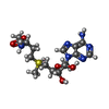  Mass: 414.480 Da / Num. of mol.: 1 / Source method: obtained synthetically / Formula: C16H26N6O5S Mass: 414.480 Da / Num. of mol.: 1 / Source method: obtained synthetically / Formula: C16H26N6O5S |
| #5: Water | ChemComp-HOH /  Mass: 18.015 Da / Num. of mol.: 163 / Source method: isolated from a natural source / Formula: H2O Mass: 18.015 Da / Num. of mol.: 163 / Source method: isolated from a natural source / Formula: H2O |
-Experimental details
-Experiment
| Experiment | Method: X-RAY DIFFRACTION / Number of used crystals: 1 |
|---|
- Sample preparation
| Crystal | Density Matthews: 2.22 Å3/Da / Density % sol: 44.61 % |
|---|---|
| Crystal grow | Temperature: 295 K / Method: vapor diffusion, hanging drop / pH: 8 Details: 16% PEG 8000, 100 mM Tris, 10 mM DTT, pH 8.0, vapor diffusion, hanging drop, temperature 295K |
-Data collection
| Diffraction | Mean temperature: 100 K |
|---|---|
| Diffraction source | Source: SYNCHROTRON / Site: APS  / Beamline: 24-ID-C / Wavelength: 0.9795 Å / Beamline: 24-ID-C / Wavelength: 0.9795 Å |
| Detector | Type: ADSC QUANTUM 315 / Detector: CCD / Date: Mar 1, 2006 |
| Radiation | Protocol: SINGLE WAVELENGTH / Monochromatic (M) / Laue (L): M / Scattering type: x-ray |
| Radiation wavelength | Wavelength: 0.9795 Å / Relative weight: 1 |
| Reflection | Resolution: 1.7→50 Å / Num. all: 35113 / Num. obs: 33418 / % possible obs: 96.3 % / Observed criterion σ(F): 0 / Observed criterion σ(I): 0 / Redundancy: 3.5 % / Biso Wilson estimate: 21.7 Å2 / Rmerge(I) obs: 0.083 / Rsym value: 0.083 / Χ2: 1.386 / Net I/σ(I): 15.374 |
| Reflection shell | Resolution: 1.7→1.76 Å / Redundancy: 2.5 % / Rmerge(I) obs: 0.328 / Mean I/σ(I) obs: 2.2 / Num. unique all: 2801 / Rsym value: 0.328 / Χ2: 0.944 / % possible all: 78 |
- Processing
| Software |
| ||||||||||||||||||||||||||||||||||||
|---|---|---|---|---|---|---|---|---|---|---|---|---|---|---|---|---|---|---|---|---|---|---|---|---|---|---|---|---|---|---|---|---|---|---|---|---|---|
| Refinement | Method to determine structure: MOLECULAR REPLACEMENT Starting model: 1I7B Resolution: 1.7→47.93 Å / Rfactor Rfree error: 0.006 / Occupancy max: 1 / Occupancy min: 1 / Data cutoff high absF: 648036 / Data cutoff low absF: 0 / Isotropic thermal model: RESTRAINED / Cross valid method: THROUGHOUT / σ(F): 0 / σ(I): 0 / Stereochemistry target values: Engh & Huber / Details: BULK SOLVENT MODEL USED
| ||||||||||||||||||||||||||||||||||||
| Solvent computation | Solvent model: FLAT MODEL / Bsol: 44.249 Å2 / ksol: 0.45 e/Å3 | ||||||||||||||||||||||||||||||||||||
| Displacement parameters | Biso max: 86.48 Å2 / Biso mean: 30.914 Å2 / Biso min: 15.39 Å2
| ||||||||||||||||||||||||||||||||||||
| Refine analyze |
| ||||||||||||||||||||||||||||||||||||
| Refinement step | Cycle: LAST / Resolution: 1.7→47.93 Å
| ||||||||||||||||||||||||||||||||||||
| Refine LS restraints |
| ||||||||||||||||||||||||||||||||||||
| LS refinement shell | Resolution: 1.7→1.81 Å / Rfactor Rfree error: 0.02 / Total num. of bins used: 6
| ||||||||||||||||||||||||||||||||||||
| Xplor file |
|
