 Movie
Movie Controller


+ Open data

- Basic information
| Entry | Database: PDB / ID: 6wwa | ||||||
|---|---|---|---|---|---|---|---|
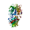
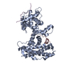
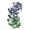
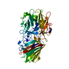
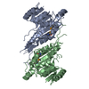
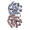
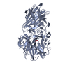
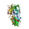
| Title | Crystal structure of human SHLD2-SHLD3-REV7 complex | ||||||
 Components Components |
| ||||||
 Keywords Keywords | NUCLEAR PROTEIN / REV7 / SHLD2 / SHLD3 | ||||||
| Function / homology |  Function and homology information Function and homology informationsomatic diversification of immunoglobulins involved in immune response / DNA damage response, signal transduction resulting in transcription / negative regulation of ubiquitin protein ligase activity / zeta DNA polymerase complex / positive regulation of isotype switching / : / JUN kinase binding / negative regulation of cell-cell adhesion mediated by cadherin / negative regulation of epithelial to mesenchymal transition / positive regulation of double-strand break repair via nonhomologous end joining ...somatic diversification of immunoglobulins involved in immune response / DNA damage response, signal transduction resulting in transcription / negative regulation of ubiquitin protein ligase activity / zeta DNA polymerase complex / positive regulation of isotype switching / : / JUN kinase binding / negative regulation of cell-cell adhesion mediated by cadherin / negative regulation of epithelial to mesenchymal transition / positive regulation of double-strand break repair via nonhomologous end joining / regulation of double-strand break repair via homologous recombination / mitotic spindle assembly checkpoint signaling / telomere maintenance in response to DNA damage / positive regulation of peptidyl-serine phosphorylation / error-prone translesion synthesis / negative regulation of double-strand break repair via homologous recombination / site of DNA damage / translesion synthesis / Translesion synthesis by REV1 / Translesion synthesis by POLK / actin filament organization / Translesion synthesis by POLI / regulation of cell growth / negative regulation of canonical Wnt signaling pathway / negative regulation of protein catabolic process / spindle / transcription corepressor activity / double-strand break repair / chromosome / actin cytoskeleton / site of double-strand break / RNA polymerase II-specific DNA-binding transcription factor binding / protein-macromolecule adaptor activity / cell division / DNA repair / positive regulation of DNA-templated transcription / chromatin / nucleolus / negative regulation of transcription by RNA polymerase II / nucleoplasm / nucleus / cytoplasm Similarity search - Function | ||||||
| Biological species |  Homo sapiens (human) Homo sapiens (human) | ||||||
| Method |  X-RAY DIFFRACTION / SYNCHROTRON / MOLECULAR REPLACEMENT / Resolution: 3.8 Å X-RAY DIFFRACTION / SYNCHROTRON / MOLECULAR REPLACEMENT / Resolution: 3.8 Å | ||||||
 Authors Authors | Xie, W. / Patel, D.J. | ||||||
 Citation Citation | Journal: Proc Natl Acad Sci U S A / Year: 2021 Title: Molecular mechanisms of assembly and TRIP13-mediated remodeling of the human Shieldin complex. Authors: Wei Xie / Shengliu Wang / Juncheng Wang / M Jason de la Cruz / Guotai Xu / Maurizio Scaltriti / Dinshaw J Patel /  Abstract: The Shieldin complex, composed of REV7, SHLD1, SHLD2, and SHLD3, protects DNA double-strand breaks (DSBs) to promote nonhomologous end joining. The AAA ATPase TRIP13 remodels Shieldin to regulate DNA ...The Shieldin complex, composed of REV7, SHLD1, SHLD2, and SHLD3, protects DNA double-strand breaks (DSBs) to promote nonhomologous end joining. The AAA ATPase TRIP13 remodels Shieldin to regulate DNA repair pathway choice. Here we report crystal structures of human SHLD3-REV7 binary and fused SHLD2-SHLD3-REV7 ternary complexes, revealing that assembly of Shieldin requires fused SHLD2-SHLD3 induced conformational heterodimerization of open (O-REV7) and closed (C-REV7) forms of REV7. We also report the cryogenic electron microscopy (cryo-EM) structures of the ATPγS-bound fused SHLD2-SHLD3-REV7-TRIP13 complexes, uncovering the principles underlying the TRIP13-mediated disassembly mechanism of the Shieldin complex. We demonstrate that the N terminus of REV7 inserts into the central channel of TRIP13, setting the stage for pulling the unfolded N-terminal peptide of C-REV7 through the central TRIP13 hexameric channel. The primary interface involves contacts between the safety-belt segment of C-REV7 and a conserved and negatively charged loop of TRIP13. This process is mediated by ATP hydrolysis-triggered rotatory motions of the TRIP13 ATPase, thereby resulting in the disassembly of the Shieldin complex. | ||||||
| History |
|
- Structure visualization
| Structure viewer | Molecule: MolmilJmol/JSmol |
|---|
- Downloads & links
-Download
| PDBx/mmCIF format | 6wwa.cif.gz | 376.9 KB | Display | PDBx/mmCIF format |
|---|---|---|---|---|
| PDB format | pdb6wwa.ent.gz | 300 KB | Display | PDB format |
| PDBx/mmJSON format | 6wwa.json.gz | Tree view | PDBx/mmJSON format | |
| Others |  Other downloads Other downloads |
-Validation report
| Arichive directory | https://data.pdbj.org/pub/pdb/validation_reports/ww/6wwaftp://data.pdbj.org/pub/pdb/validation_reports/ww/6wwa | HTTPS FTP |
|---|
-Related structure data
| Related structure data |  6ww9C 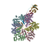 7l9pC 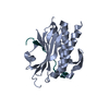 3abdS S: Starting model for refinement C: citing same article ( |
|---|---|
| Similar structure data |


-Links
 PDBj
PDBj


- Assembly
| Deposited unit | 
| ||||||||||||
|---|---|---|---|---|---|---|---|---|---|---|---|---|---|
| 1 | 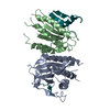
| ||||||||||||
| 2 | 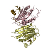
| ||||||||||||
| Unit cell |
|
-Components
| #1: Protein | Mass: 24323.348 Da / Num. of mol.: 4 Source method: isolated from a genetically manipulated source Source: (gene. exp.) Homo sapiens (human) / Gene: MAD2L2, MAD2B, REV7 / Production host:  #2: Protein | Mass: 10678.139 Da / Num. of mol.: 2 Source method: isolated from a genetically manipulated source Source: (gene. exp.) Homo sapiens (human) / Gene: SHLD2, FAM35A, RINN2, SHLD3, FLJ26957, RINN1 / Production host: |
|---|
-Experimental details
-Experiment
| Experiment | Method: X-RAY DIFFRACTION / Number of used crystals: 1 |
|---|
- Sample preparation
| Crystal | Density Matthews: 6.69 Å3/Da / Density % sol: 81.6 % |
|---|---|
| Crystal grow | Temperature: 293 K / Method: vapor diffusion, hanging drop / pH: 5.6 / Details: 0.1 M Na acetate pH 5.6, 1.5 M Na formate |
-Data collection
| Diffraction | Mean temperature: 100 K / Serial crystal experiment: N |
|---|---|
| Diffraction source | Source: SYNCHROTRON / Site: APS / Beamline: 24-ID-C / Wavelength: 0.9792 Å |
| Detector | Type: PSI PILATUS 6M / Detector: PIXEL / Date: Dec 16, 2019 |
| Radiation | Protocol: SINGLE WAVELENGTH / Monochromatic (M) / Laue (L): M / Scattering type: x-ray |
| Radiation wavelength | Wavelength: 0.9792 Å / Relative weight: 1 |
| Reflection | Resolution: 3.8→40 Å / Num. obs: 30856 / % possible obs: 99.74 % / Redundancy: 35.8 % / Biso Wilson estimate: 153.27 Å2 / CC1/2: 0.999 / CC star: 1 / Rmerge(I) obs: 0.4031 / Rpim(I) all: 0.06797 / Rrim(I) all: 0.4089 / Net I/σ(I): 10.3 |
| Reflection shell | Resolution: 3.8→3.936 Å / Rmerge(I) obs: 4.971 / Mean I/σ(I) obs: 0.86 / Num. unique obs: 3027 / CC1/2: 0.472 / CC star: 0.801 / Rpim(I) all: 0.8316 / Rrim(I) all: 5.04 |
- Processing
| Software |
| ||||||||||||||||||||||||||||||||||||||||||||||||||||||||||||||||||||||||||||||||||||
|---|---|---|---|---|---|---|---|---|---|---|---|---|---|---|---|---|---|---|---|---|---|---|---|---|---|---|---|---|---|---|---|---|---|---|---|---|---|---|---|---|---|---|---|---|---|---|---|---|---|---|---|---|---|---|---|---|---|---|---|---|---|---|---|---|---|---|---|---|---|---|---|---|---|---|---|---|---|---|---|---|---|---|---|---|---|
| Refinement | Method to determine structure: MOLECULAR REPLACEMENT Starting model: 3ABD Resolution: 3.8→39.66 Å / SU ML: 0.5072 / Cross valid method: FREE R-VALUE / σ(F): 1.34 / Phase error: 29.5099 Stereochemistry target values: GeoStd + Monomer Library + CDL v1.2
| ||||||||||||||||||||||||||||||||||||||||||||||||||||||||||||||||||||||||||||||||||||
| Solvent computation | Shrinkage radii: 0.9 Å / VDW probe radii: 1.11 Å / Solvent model: FLAT BULK SOLVENT MODEL | ||||||||||||||||||||||||||||||||||||||||||||||||||||||||||||||||||||||||||||||||||||
| Displacement parameters | Biso mean: 189.45 Å2 | ||||||||||||||||||||||||||||||||||||||||||||||||||||||||||||||||||||||||||||||||||||
| Refinement step | Cycle: LAST / Resolution: 3.8→39.66 Å
| ||||||||||||||||||||||||||||||||||||||||||||||||||||||||||||||||||||||||||||||||||||
| Refine LS restraints |
| ||||||||||||||||||||||||||||||||||||||||||||||||||||||||||||||||||||||||||||||||||||
| LS refinement shell |
| ||||||||||||||||||||||||||||||||||||||||||||||||||||||||||||||||||||||||||||||||||||
| Refinement TLS params. | Method: refined / Origin x: 109.905339802 Å / Origin y: 14.6069753103 Å / Origin z: 133.372128478 Å
| ||||||||||||||||||||||||||||||||||||||||||||||||||||||||||||||||||||||||||||||||||||
| Refinement TLS group | Selection details: all |
