Movie
Movie Controller


+ Open data

- Basic information
| Entry | Database: EMDB / ID: EMD-23244 | |||||||||
|---|---|---|---|---|---|---|---|---|---|---|
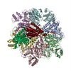
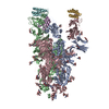
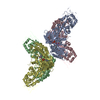
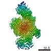
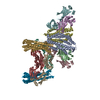
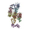
| Title | Structure of human SHLD2-SHLD3-REV7-TRIP13(E253Q) complex | |||||||||
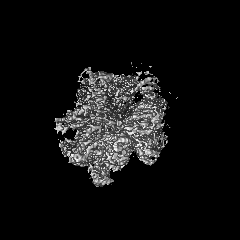
 Map data Map data | composite map | |||||||||
 Sample Sample |
| |||||||||
 Keywords Keywords | REV7 / SHLD2 / SHLD3 / TRIP13 / NUCLEAR PROTEIN | |||||||||
| Function / homology |  Function and homology information Function and homology informationmeiotic recombination checkpoint signaling / somatic diversification of immunoglobulins involved in immune response / DNA damage response, signal transduction resulting in transcription / negative regulation of ubiquitin protein ligase activity / zeta DNA polymerase complex / synaptonemal complex assembly / positive regulation of isotype switching / : / JUN kinase binding / negative regulation of cell-cell adhesion mediated by cadherin ...meiotic recombination checkpoint signaling / somatic diversification of immunoglobulins involved in immune response / DNA damage response, signal transduction resulting in transcription / negative regulation of ubiquitin protein ligase activity / zeta DNA polymerase complex / synaptonemal complex assembly / positive regulation of isotype switching / : / JUN kinase binding / negative regulation of cell-cell adhesion mediated by cadherin / reciprocal meiotic recombination / oogenesis / negative regulation of epithelial to mesenchymal transition / positive regulation of double-strand break repair via nonhomologous end joining / mitotic spindle assembly checkpoint signaling / regulation of double-strand break repair via homologous recombination / telomere maintenance in response to DNA damage / positive regulation of peptidyl-serine phosphorylation / error-prone translesion synthesis / negative regulation of double-strand break repair via homologous recombination / site of DNA damage / translesion synthesis / male germ cell nucleus / actin filament organization / Translesion synthesis by REV1 / Translesion synthesis by POLK / Translesion synthesis by POLI / transcription coregulator activity / negative regulation of canonical Wnt signaling pathway / regulation of cell growth / negative regulation of protein catabolic process / spindle / transcription corepressor activity / actin cytoskeleton / transcription by RNA polymerase II / double-strand break repair / chromosome / site of double-strand break / spermatogenesis / RNA polymerase II-specific DNA-binding transcription factor binding / protein-macromolecule adaptor activity / cell division / DNA repair / positive regulation of DNA-templated transcription / chromatin / negative regulation of transcription by RNA polymerase II / ATP hydrolysis activity / DNA-templated transcription / nucleoplasm / ATP binding / identical protein binding / nucleus / cytoplasm Similarity search - Function | |||||||||
| Biological species |  Homo sapiens (human) Homo sapiens (human) | |||||||||
| Method | single particle reconstruction / cryo EM / Resolution: 3.6 Å | |||||||||
 Authors Authors | Xie W / Patel DJ | |||||||||
 Citation Citation | Journal: Proc Natl Acad Sci U S A / Year: 2021 Title: Molecular mechanisms of assembly and TRIP13-mediated remodeling of the human Shieldin complex. Authors: Wei Xie / Shengliu Wang / Juncheng Wang / M Jason de la Cruz / Guotai Xu / Maurizio Scaltriti / Dinshaw J Patel /  Abstract: The Shieldin complex, composed of REV7, SHLD1, SHLD2, and SHLD3, protects DNA double-strand breaks (DSBs) to promote nonhomologous end joining. The AAA ATPase TRIP13 remodels Shieldin to regulate DNA ...The Shieldin complex, composed of REV7, SHLD1, SHLD2, and SHLD3, protects DNA double-strand breaks (DSBs) to promote nonhomologous end joining. The AAA ATPase TRIP13 remodels Shieldin to regulate DNA repair pathway choice. Here we report crystal structures of human SHLD3-REV7 binary and fused SHLD2-SHLD3-REV7 ternary complexes, revealing that assembly of Shieldin requires fused SHLD2-SHLD3 induced conformational heterodimerization of open (O-REV7) and closed (C-REV7) forms of REV7. We also report the cryogenic electron microscopy (cryo-EM) structures of the ATPγS-bound fused SHLD2-SHLD3-REV7-TRIP13 complexes, uncovering the principles underlying the TRIP13-mediated disassembly mechanism of the Shieldin complex. We demonstrate that the N terminus of REV7 inserts into the central channel of TRIP13, setting the stage for pulling the unfolded N-terminal peptide of C-REV7 through the central TRIP13 hexameric channel. The primary interface involves contacts between the safety-belt segment of C-REV7 and a conserved and negatively charged loop of TRIP13. This process is mediated by ATP hydrolysis-triggered rotatory motions of the TRIP13 ATPase, thereby resulting in the disassembly of the Shieldin complex. | |||||||||
| History |
|
- Structure visualization
| Movie |
Movie viewer |
|---|---|
| Structure viewer | EM map: SurfViewMolmilJmol/JSmol |
| Supplemental images |

- Downloads & links
-EMDB archive
| Map data | emd_23244.map.gz | 49.3 MB | EMDB map data format | |
|---|---|---|---|---|
| Header (meta data) | emd-23244-v30.xmlemd-23244.xml | 15.8 KB 15.8 KB | Display Display | EMDB header |
| Images | 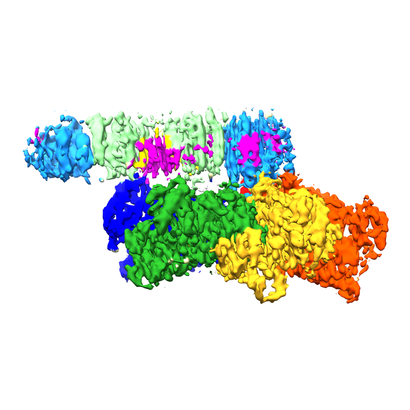 emd_23244.png emd_23244.png | 314.2 KB | ||
| Filedesc metadata | emd-23244.cif.gz | 6.4 KB | ||
| Archive directory |  http://ftp.pdbj.org/pub/emdb/structures/EMD-23244ftp://ftp.pdbj.org/pub/emdb/structures/EMD-23244 http://ftp.pdbj.org/pub/emdb/structures/EMD-23244ftp://ftp.pdbj.org/pub/emdb/structures/EMD-23244 | HTTPS FTP |
-Related structure data
| Related structure data |  7l9pMC  6ww9C  6wwaC M: atomic model generated by this map C: citing same article ( |
|---|---|
| Similar structure data |

-Links
| EMDB pages | EMDB (EBI/PDBe) / EMDataResource |
|---|---|
| Related items in Molecule of the Month |


-Map
| File | Download / File: emd_23244.map.gz / Format: CCP4 / Size: 52.7 MB / Type: IMAGE STORED AS FLOATING POINT NUMBER (4 BYTES) | ||||||||||||||||||||||||||||||||||||||||||||||||||||||||||||||||||||
|---|---|---|---|---|---|---|---|---|---|---|---|---|---|---|---|---|---|---|---|---|---|---|---|---|---|---|---|---|---|---|---|---|---|---|---|---|---|---|---|---|---|---|---|---|---|---|---|---|---|---|---|---|---|---|---|---|---|---|---|---|---|---|---|---|---|---|---|---|---|
| Annotation | composite map | ||||||||||||||||||||||||||||||||||||||||||||||||||||||||||||||||||||
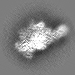
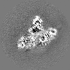
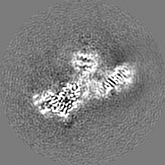
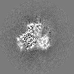
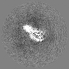
| Projections & slices | Image control
Images are generated by Spider. | ||||||||||||||||||||||||||||||||||||||||||||||||||||||||||||||||||||
| Voxel size | X=Y=Z: 1.064 Å | ||||||||||||||||||||||||||||||||||||||||||||||||||||||||||||||||||||
| Density |
| ||||||||||||||||||||||||||||||||||||||||||||||||||||||||||||||||||||
| Symmetry | Space group: 1 | ||||||||||||||||||||||||||||||||||||||||||||||||||||||||||||||||||||
| Details | EMDB XML:
CCP4 map header:
| ||||||||||||||||||||||||||||||||||||||||||||||||||||||||||||||||||||
 X (Sec.)
 Y (Row.)
 Z (Col.)


-Supplemental data
- Sample components
-Entire : SHLD2.3-REV7(4)-TRIP13(E253Q) complex with ATP-gamma-S
| Entire | Name: SHLD2.3-REV7(4)-TRIP13(E253Q) complex with ATP-gamma-S |
|---|---|
| Components |
|
-Supramolecule #1: SHLD2.3-REV7(4)-TRIP13(E253Q) complex with ATP-gamma-S
| Supramolecule | Name: SHLD2.3-REV7(4)-TRIP13(E253Q) complex with ATP-gamma-S type: complex / ID: 1 / Parent: 0 / Macromolecule list: #1-#3 |
|---|---|
| Source (natural) | Organism: Homo sapiens (human) |
-Macromolecule #1: Pachytene checkpoint protein 2 homolog
| Macromolecule | Name: Pachytene checkpoint protein 2 homolog / type: protein_or_peptide / ID: 1 / Number of copies: 6 / Enantiomer: LEVO |
|---|---|
| Source (natural) | Organism: Homo sapiens (human) |
| Molecular weight | Theoretical: 48.562547 KDa |
| Recombinant expression | Organism:  |
| Sequence | String: SDEAVGDLKQ ALPCVAESPT VHVEVHQRGS STAKKEDINL SVRKLLNRHN IVFGDYTWTE FDEPFLTRNV QSVSIIDTEL KVKDSQPID LSACTVALHI FQLNEDGPSS ENLEEETENI IAANHWVLPA AEFHGLWDSL VYDVEVKSHL LDYVMTTLLF S DKNVNSNL ...String: SDEAVGDLKQ ALPCVAESPT VHVEVHQRGS STAKKEDINL SVRKLLNRHN IVFGDYTWTE FDEPFLTRNV QSVSIIDTEL KVKDSQPID LSACTVALHI FQLNEDGPSS ENLEEETENI IAANHWVLPA AEFHGLWDSL VYDVEVKSHL LDYVMTTLLF S DKNVNSNL ITWNRVVLLH GPPGTGKTSL CKALAQKLTI RLSSRYRYGQ LIEINSHSLF SKWFSESGKL VTKMFQKIQD LI DDKDALV FVLIDQVESL TAARNACRAG TEPSDAIRVV NAVLTQIDQI KRHSNVVILT TSNITEKIDV AFVDRADIKQ YIG PPSAAA IFKIYLSCLE ELMKCQIIYP RQQLLTLREL EMIGFIENNV SKLSLLLNDI SRKSEGLSGR VLRKLPFLAH ALYV QAPTV TIEGFLQALS LAVDKQFEER KKLAAYI UniProtKB: Pachytene checkpoint protein 2 homolog |
-Macromolecule #2: Mitotic spindle assembly checkpoint protein MAD2B
| Macromolecule | Name: Mitotic spindle assembly checkpoint protein MAD2B / type: protein_or_peptide / ID: 2 / Details: closed form / Number of copies: 4 / Enantiomer: LEVO |
|---|---|
| Source (natural) | Organism: Homo sapiens (human) |
| Molecular weight | Theoretical: 24.323348 KDa |
| Recombinant expression | Organism: |
| Sequence | String: STTLTRQDLN FGQVVADVLC EFLEVAVHLI LYVREVYPVG IFQKRKKYNV PVQMSCHPEL NQYIQDTLHC VKPLLEKNDV EKVVVVILD KEHRPVEKFV FEITQPPLLS ISSDSLLSHV EQLLRAFILK ISVCDAVLDH NPPGCTFTVL VHTREAATRN M EKIQVIKD ...String: STTLTRQDLN FGQVVADVLC EFLEVAVHLI LYVREVYPVG IFQKRKKYNV PVQMSCHPEL NQYIQDTLHC VKPLLEKNDV EKVVVVILD KEHRPVEKFV FEITQPPLLS ISSDSLLSHV EQLLRAFILK ISVCDAVLDH NPPGCTFTVL VHTREAATRN M EKIQVIKD FPWILADEQD VHMHDPRLIP LKTMTSDILK MQLYVEERAH KGS UniProtKB: Mitotic spindle assembly checkpoint protein MAD2B |
-Macromolecule #3: Shieldin complex subunit 2, Shieldin complex subunit 3 chimera
| Macromolecule | Name: Shieldin complex subunit 2, Shieldin complex subunit 3 chimera type: protein_or_peptide / ID: 3 / Number of copies: 2 / Enantiomer: LEVO |
|---|---|
| Source (natural) | Organism: Homo sapiens (human) |
| Molecular weight | Theoretical: 10.678139 KDa |
| Recombinant expression | Organism: |
| Sequence | String: MSQVHIFWGA PIAPLKGSGS GSGSGSGSGS GSTTEVILHY RPCESDPTQL PKIAEKAIQD FPTRPLSRFI PWFPYDGSKL PLRPKRSPP ASREEIMATL UniProtKB: Shieldin complex subunit 2, Shieldin complex subunit 3 |
-Macromolecule #4: PHOSPHOTHIOPHOSPHORIC ACID-ADENYLATE ESTER
| Macromolecule | Name: PHOSPHOTHIOPHOSPHORIC ACID-ADENYLATE ESTER / type: ligand / ID: 4 / Number of copies: 5 / Formula: AGS |
|---|---|
| Molecular weight | Theoretical: 523.247 Da |
| Chemical component information |  ChemComp-AGS: |
-Experimental details
-Structure determination
| Method | cryo EM |
|---|---|
 Processing Processing | single particle reconstruction |
| Aggregation state | particle |
-Sample preparation
| Concentration | 0.3 mg/mL |
|---|---|
| Buffer | pH: 7.3 Details: 20 mM HEPES, pH 7.3, 300 mM NaCl, 5 mM MgCl2, 0.1 mM ATP-gamma-S, 1 mM DTT |
| Grid | Model: UltrAuFoil R1.2/1.3 / Material: GOLD / Pretreatment - Type: GLOW DISCHARGE / Pretreatment - Time: 60 sec. |
| Vitrification | Cryogen name: ETHANE / Chamber humidity: 100 % / Chamber temperature: 277 K / Instrument: FEI VITROBOT MARK IV / Details: 1.5-second blot, blot force of 0. |
- Electron microscopy
| Microscope | FEI TITAN KRIOS |
|---|---|
| Image recording | Film or detector model: GATAN K3 BIOQUANTUM (6k x 4k) / Number grids imaged: 1 / Number real images: 40 / Average exposure time: 0.075 sec. / Average electron dose: 53.0 e/Å2 |
| Electron beam | Acceleration voltage: 300 kV / Electron source:  FIELD EMISSION GUN FIELD EMISSION GUN |
| Electron optics | Illumination mode: FLOOD BEAM / Imaging mode: BRIGHT FIELD / Nominal defocus max: -2.5 µm / Nominal defocus min: -1.0 µm |
| Sample stage | Specimen holder model: FEI TITAN KRIOS AUTOGRID HOLDER / Cooling holder cryogen: NITROGEN |
| Experimental equipment |  Model: Titan Krios / Image courtesy: FEI Company |