 Movie
Movie Controller

[English] 日本語
 Yorodumi
- PDB-6f0x: Cryo-EM structure of TRIP13 in complex with ATP gamma S, p31comet... -
+ Open data

- Basic information
| Entry | Database: PDB / ID: 6f0x | ||||||||||||||||||||||||||||||||||||||||||||||||||||||||||||||||||||||||||||||||||||||||||||||||||||||
|---|---|---|---|---|---|---|---|---|---|---|---|---|---|---|---|---|---|---|---|---|---|---|---|---|---|---|---|---|---|---|---|---|---|---|---|---|---|---|---|---|---|---|---|---|---|---|---|---|---|---|---|---|---|---|---|---|---|---|---|---|---|---|---|---|---|---|---|---|---|---|---|---|---|---|---|---|---|---|---|---|---|---|---|---|---|---|---|---|---|---|---|---|---|---|---|---|---|---|---|---|---|---|---|
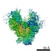
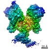
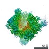
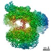
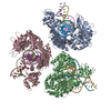
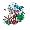
| Title | Cryo-EM structure of TRIP13 in complex with ATP gamma S, p31comet, C-Mad2 and Cdc20 | ||||||||||||||||||||||||||||||||||||||||||||||||||||||||||||||||||||||||||||||||||||||||||||||||||||||
 Components Components |
| ||||||||||||||||||||||||||||||||||||||||||||||||||||||||||||||||||||||||||||||||||||||||||||||||||||||
 Keywords Keywords | CELL CYCLE / AAA+ ATPase / remodeller / spindle assembly checkpoint (SAC) / mitosis / chromosome segregation | ||||||||||||||||||||||||||||||||||||||||||||||||||||||||||||||||||||||||||||||||||||||||||||||||||||||
| Function / homology |  Function and homology information Function and homology informationdeactivation of mitotic spindle assembly checkpoint / mitotic spindle assembly checkpoint MAD1-MAD2 complex / metaphase/anaphase transition of cell cycle / mitotic checkpoint complex / metaphase/anaphase transition of meiosis I / meiotic recombination checkpoint signaling / Inhibition of the proteolytic activity of APC/C required for the onset of anaphase by mitotic spindle checkpoint components / positive regulation of ubiquitin protein ligase activity / negative regulation of ubiquitin protein ligase activity / positive regulation of anaphase-promoting complex-dependent catabolic process ...deactivation of mitotic spindle assembly checkpoint / mitotic spindle assembly checkpoint MAD1-MAD2 complex / metaphase/anaphase transition of cell cycle / mitotic checkpoint complex / metaphase/anaphase transition of meiosis I / meiotic recombination checkpoint signaling / Inhibition of the proteolytic activity of APC/C required for the onset of anaphase by mitotic spindle checkpoint components / positive regulation of ubiquitin protein ligase activity / negative regulation of ubiquitin protein ligase activity / positive regulation of anaphase-promoting complex-dependent catabolic process / positive regulation of synapse maturation / synaptonemal complex assembly / Conversion from APC/C:Cdc20 to APC/C:Cdh1 in late anaphase / Inactivation of APC/C via direct inhibition of the APC/C complex / APC/C:Cdc20 mediated degradation of mitotic proteins / regulation of dendrite development / anaphase-promoting complex / Phosphorylation of Emi1 / anaphase-promoting complex-dependent catabolic process / regulation of meiotic cell cycle / anaphase-promoting complex binding / regulation of exit from mitosis / oocyte maturation / nuclear pore nuclear basket / positive regulation of synaptic plasticity / negative regulation of mitotic cell cycle / positive regulation of mitotic cell cycle spindle assembly checkpoint / positive regulation of mitotic metaphase/anaphase transition / female meiosis I / ubiquitin ligase activator activity / reciprocal meiotic recombination / oogenesis / mitotic spindle assembly checkpoint signaling / spermatid development / Regulation of APC/C activators between G1/S and early anaphase / mitotic sister chromatid segregation / male meiosis I / ubiquitin-like ligase-substrate adaptor activity / Amplification of signal from unattached kinetochores via a MAD2 inhibitory signal / regulation of mitotic cell cycle / Mitotic Prometaphase / EML4 and NUDC in mitotic spindle formation / APC/C:Cdc20 mediated degradation of Cyclin B / APC-Cdc20 mediated degradation of Nek2A / male germ cell nucleus / Resolution of Sister Chromatid Cohesion / transcription coregulator activity / APC/C:Cdc20 mediated degradation of Securin / SCF-beta-TrCP mediated degradation of Emi1 / Cdc20:Phospho-APC/C mediated degradation of Cyclin A / RHO GTPases Activate Formins / negative regulation of protein catabolic process / APC/C:Cdh1 mediated degradation of Cdc20 and other APC/C:Cdh1 targeted proteins in late mitosis/early G1 / kinetochore / spindle / histone deacetylase binding / spindle pole / mitotic spindle / Separation of Sister Chromatids / double-strand break repair / nervous system development / Antigen processing: Ubiquitination & Proteasome degradation / chromosome / transcription by RNA polymerase II / nuclear membrane / spermatogenesis / cell differentiation / Ub-specific processing proteases / protein ubiquitination / cell division / centrosome / positive regulation of cell population proliferation / nucleolus / perinuclear region of cytoplasm / protein homodimerization activity / ATP hydrolysis activity / nucleoplasm / ATP binding / identical protein binding / nucleus / cytosol / cytoplasm Similarity search - Function | ||||||||||||||||||||||||||||||||||||||||||||||||||||||||||||||||||||||||||||||||||||||||||||||||||||||
| Biological species |  Homo sapiens (human) Homo sapiens (human) | ||||||||||||||||||||||||||||||||||||||||||||||||||||||||||||||||||||||||||||||||||||||||||||||||||||||
| Method | ELECTRON MICROSCOPY / single particle reconstruction / cryo EM / Resolution: 4.6 Å | ||||||||||||||||||||||||||||||||||||||||||||||||||||||||||||||||||||||||||||||||||||||||||||||||||||||
 Authors Authors | Alfieri, C. / Chang, L. / Barford, D. | ||||||||||||||||||||||||||||||||||||||||||||||||||||||||||||||||||||||||||||||||||||||||||||||||||||||
| Funding support |  United Kingdom, 1items United Kingdom, 1items
| ||||||||||||||||||||||||||||||||||||||||||||||||||||||||||||||||||||||||||||||||||||||||||||||||||||||
 Citation Citation | Journal: Nature / Year: 2018 Title: Mechanism for remodelling of the cell cycle checkpoint protein MAD2 by the ATPase TRIP13. Authors: Claudio Alfieri / Leifu Chang / David Barford /  Abstract: The maintenance of genome stability during mitosis is coordinated by the spindle assembly checkpoint (SAC) through its effector the mitotic checkpoint complex (MCC), an inhibitor of the anaphase- ...The maintenance of genome stability during mitosis is coordinated by the spindle assembly checkpoint (SAC) through its effector the mitotic checkpoint complex (MCC), an inhibitor of the anaphase-promoting complex (APC/C, also known as the cyclosome). Unattached kinetochores control MCC assembly by catalysing a change in the topology of the β-sheet of MAD2 (an MCC subunit), thereby generating the active closed MAD2 (C-MAD2) conformer. Disassembly of free MCC, which is required for SAC inactivation and chromosome segregation, is an ATP-dependent process driven by the AAA+ ATPase TRIP13. In combination with p31, an SAC antagonist, TRIP13 remodels C-MAD2 into inactive open MAD2 (O-MAD2). Here, we present a mechanism that explains how TRIP13-p31 disassembles the MCC. Cryo-electron microscopy structures of the TRIP13-p31-C-MAD2-CDC20 complex reveal that p31 recruits C-MAD2 to a defined site on the TRIP13 hexameric ring, positioning the N terminus of C-MAD2 (MAD2) to insert into the axial pore of TRIP13 and distorting the TRIP13 ring to initiate remodelling. Molecular modelling suggests that by gripping MAD2 within its axial pore, TRIP13 couples sequential ATP-driven translocation of its hexameric ring along MAD2 to push upwards on, and simultaneously rotate, the globular domains of the p31-C-MAD2 complex. This unwinds a region of the αA helix of C-MAD2 that is required to stabilize the C-MAD2 β-sheet, thus destabilizing C-MAD2 in favour of O-MAD2 and dissociating MAD2 from p31. Our study provides insights into how specific substrates are recruited to AAA+ ATPases through adaptor proteins and suggests a model of how translocation through the axial pore of AAA+ ATPases is coupled to protein remodelling. | ||||||||||||||||||||||||||||||||||||||||||||||||||||||||||||||||||||||||||||||||||||||||||||||||||||||
| History |
|
- Structure visualization
| Movie |
Movie viewer |
|---|---|
| Structure viewer | Molecule: MolmilJmol/JSmol |
- Downloads & links
-Download
| PDBx/mmCIF format | 6f0x.cif.gz | 470.8 KB | Display | PDBx/mmCIF format |
|---|---|---|---|---|
| PDB format | pdb6f0x.ent.gz | 377.5 KB | Display | PDB format |
| PDBx/mmJSON format | 6f0x.json.gz | Tree view | PDBx/mmJSON format | |
| Others |  Other downloads Other downloads |
-Validation report
| Arichive directory | https://data.pdbj.org/pub/pdb/validation_reports/f0/6f0xftp://data.pdbj.org/pub/pdb/validation_reports/f0/6f0x | HTTPS FTP |
|---|
-Related structure data
| Related structure data |  4166MC M: map data used to model this data C: citing same article ( |
|---|---|
| Similar structure data |


-Links
 PDBj
PDBj


- Assembly
| Deposited unit | 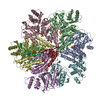
|
|---|---|
| 1 |
|
-Components
| #1: Protein | Mass: 48606.664 Da / Num. of mol.: 6 Source method: isolated from a genetically manipulated source Source: (gene. exp.) Homo sapiens (human) / Gene: TRIP13, PCH2 / Production host:  #2: Protein | | Mass: 31086.646 Da / Num. of mol.: 1 Source method: isolated from a genetically manipulated source Source: (gene. exp.) Homo sapiens (human) / Production host: #3: Protein | | Mass: 54796.508 Da / Num. of mol.: 1 Source method: isolated from a genetically manipulated source Source: (gene. exp.) Homo sapiens (human) / Gene: CDC20 / Production host:  Trichoplusia ni (cabbage looper) / References: UniProt: Q12834 Trichoplusia ni (cabbage looper) / References: UniProt: Q12834#4: Protein | | Mass: 23533.883 Da / Num. of mol.: 1 Source method: isolated from a genetically manipulated source Source: (gene. exp.) Homo sapiens (human) / Gene: MAD2L1, MAD2 / Production host: Trichoplusia ni (cabbage looper) / References: UniProt: Q13257#5: Chemical | ChemComp-AGS /   Mass: 523.247 Da / Num. of mol.: 5 / Source method: obtained synthetically / Formula: C10H16N5O12P3S / Comment: ATP-gamma-S, energy-carrying molecule analogue*YM Mass: 523.247 Da / Num. of mol.: 5 / Source method: obtained synthetically / Formula: C10H16N5O12P3S / Comment: ATP-gamma-S, energy-carrying molecule analogue*YMHas protein modification | Y | |
|---|
-Experimental details
-Experiment
| Experiment | Method: ELECTRON MICROSCOPY |
|---|---|
| EM experiment | Aggregation state: PARTICLE / 3D reconstruction method: single particle reconstruction |
- Sample preparation
| Component |
| ||||||||||||||||||||||||
|---|---|---|---|---|---|---|---|---|---|---|---|---|---|---|---|---|---|---|---|---|---|---|---|---|---|
| Molecular weight | Value: 400 kDa/nm / Experimental value: NO | ||||||||||||||||||||||||
| Source (natural) |
| ||||||||||||||||||||||||
| Source (recombinant) |
| ||||||||||||||||||||||||
| Buffer solution | pH: 7.5 | ||||||||||||||||||||||||
| Specimen | Conc.: 0.3 mg/ml / Embedding applied: NO / Shadowing applied: NO / Staining applied: NO / Vitrification applied: YES | ||||||||||||||||||||||||
| Vitrification | Cryogen name: ETHANE |
- Electron microscopy imaging
| Experimental equipment |  Model: Titan Krios / Image courtesy: FEI Company |
|---|---|
| Microscopy | Model: FEI TITAN KRIOS |
| Electron gun | Electron source:  FIELD EMISSION GUN / Accelerating voltage: 300 kV / Illumination mode: FLOOD BEAM FIELD EMISSION GUN / Accelerating voltage: 300 kV / Illumination mode: FLOOD BEAM |
| Electron lens | Mode: BRIGHT FIELD |
| Image recording | Electron dose: 40 e/Å2 / Film or detector model: GATAN K2 SUMMIT (4k x 4k) |
- Processing
| Software | Name: PHENIX / Version: dev_2919: / Classification: refinement |
|---|---|
| EM software | Name: RELION / Version: 2.1-beta-1 / Category: 3D reconstruction |
| CTF correction | Type: PHASE FLIPPING AND AMPLITUDE CORRECTION |
| Symmetry | Point symmetry: C1 (asymmetric) |
| 3D reconstruction | Resolution: 4.6 Å / Resolution method: FSC 0.143 CUT-OFF / Num. of particles: 354157 / Symmetry type: POINT |
