 Movie
Movie Controller

[English] 日本語
 Yorodumi
- PDB-5z5d: Crystal structure of a thermostable glycoside hydrolase family 43... -
+ Open data

- Basic information
| Entry | Database: PDB / ID: 5z5d | ||||||
|---|---|---|---|---|---|---|---|
| Title | Crystal structure of a thermostable glycoside hydrolase family 43 {beta}-1,4-xylosidase from Geobacillus thermoleovorans IT-08 | ||||||
 Components Components | Beta-xylosidase | ||||||
 Keywords Keywords | HYDROLASE / Xylanolytic / GH43 / oligosaccharide / saccharification | ||||||
| Function / homology |  Function and homology information Function and homology informationxylan 1,4-beta-xylosidase / xylan 1,4-beta-xylosidase activity / carbohydrate metabolic process / metal ion binding Similarity search - Function | ||||||
| Biological species |  Geobacillus thermoleovorans (bacteria) Geobacillus thermoleovorans (bacteria) | ||||||
| Method |  X-RAY DIFFRACTION / SYNCHROTRON / MOLECULAR REPLACEMENT / Resolution: 1.7 Å X-RAY DIFFRACTION / SYNCHROTRON / MOLECULAR REPLACEMENT / Resolution: 1.7 Å | ||||||
 Authors Authors | Rohman, A. / van Oosterwijk, N. / Puspaningsih, N.N.T. / Dijkstra, B.W. | ||||||
 Citation Citation | Journal: PLoS ONE / Year: 2018 Title: Structural basis of product inhibition by arabinose and xylose of the thermostable GH43 beta-1,4-xylosidase from Geobacillus thermoleovorans IT-08. Authors: Rohman, A. / van Oosterwijk, N. / Puspaningsih, N.N.T. / Dijkstra, B.W. #1: Journal: Acta Crystallogr. Sect. F Struct. Biol. Cryst. Commun. Year: 2007 Title: Purification, crystallization and preliminary X-ray analysis of a thermostable glycoside hydrolase family 43 beta-xylosidase from Geobacillus thermoleovorans IT-08. Authors: Rohman, A. / van Oosterwijk, N. / Kralj, S. / Dijkhuizen, L. / Dijkstra, B.W. / Puspaningsih, N.N. | ||||||
| History |
|
- Structure visualization
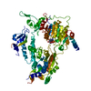
| Structure viewer | Molecule: MolmilJmol/JSmol |
|---|
- Downloads & links
-Download
| PDBx/mmCIF format | 5z5d.cif.gz | 132.7 KB | Display | PDBx/mmCIF format |
|---|---|---|---|---|
| PDB format | pdb5z5d.ent.gz | 99.3 KB | Display | PDB format |
| PDBx/mmJSON format | 5z5d.json.gz | Tree view | PDBx/mmJSON format | |
| Others |  Other downloads Other downloads |
-Validation report
| Arichive directory | https://data.pdbj.org/pub/pdb/validation_reports/z5/5z5dftp://data.pdbj.org/pub/pdb/validation_reports/z5/5z5d | HTTPS FTP |
|---|
-Related structure data
| Related structure data |  5z5fC  5z5hC  5z5iC  1yifS S: Starting model for refinement C: citing same article ( |
|---|---|
| Similar structure data |


-Links
 PDBj
PDBj- Assembly
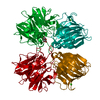
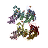
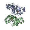
| Deposited unit | 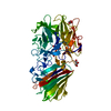
| ||||||||
|---|---|---|---|---|---|---|---|---|---|
| 1 | 
| ||||||||
| Unit cell |
|
-Components
| #1: Protein | Mass: 61603.324 Da / Num. of mol.: 1 Source method: isolated from a genetically manipulated source Source: (gene. exp.) Geobacillus thermoleovorans (bacteria) / Gene: xyl / Production host: | ||
|---|---|---|---|
| #2: Chemical | ChemComp-CA /   Mass: 40.078 Da / Num. of mol.: 1 / Source method: obtained synthetically / Formula: Ca Mass: 40.078 Da / Num. of mol.: 1 / Source method: obtained synthetically / Formula: Ca | ||
| #3: Chemical | ChemComp-GOL /   Mass: 92.094 Da / Num. of mol.: 5 / Source method: obtained synthetically / Formula: C3H8O3 Mass: 92.094 Da / Num. of mol.: 5 / Source method: obtained synthetically / Formula: C3H8O3#4: Water | ChemComp-HOH / |  Mass: 18.015 Da / Num. of mol.: 570 / Source method: isolated from a natural source / Formula: H2O Mass: 18.015 Da / Num. of mol.: 570 / Source method: isolated from a natural source / Formula: H2O |
-Experimental details
-Experiment
| Experiment | Method: X-RAY DIFFRACTION / Number of used crystals: 1 |
|---|
- Sample preparation
| Crystal | Density Matthews: 2.29 Å3/Da / Density % sol: 46.35 % |
|---|---|
| Crystal grow | Temperature: 283 K / Method: vapor diffusion, hanging drop / pH: 7 Details: 0.1M HEPES (N-(2-hydroxyethyl)piperazine-N-(2-ethanesulfonic acid)), pH 7.0, 5% (w/v) PEG 6000 |
-Data collection
| Diffraction | Mean temperature: 100 K | |||||||||||||||||||||||||||||||||||||||||||||||||||||||||||||||||||||||||||||||||||||||||||||||||||
|---|---|---|---|---|---|---|---|---|---|---|---|---|---|---|---|---|---|---|---|---|---|---|---|---|---|---|---|---|---|---|---|---|---|---|---|---|---|---|---|---|---|---|---|---|---|---|---|---|---|---|---|---|---|---|---|---|---|---|---|---|---|---|---|---|---|---|---|---|---|---|---|---|---|---|---|---|---|---|---|---|---|---|---|---|---|---|---|---|---|---|---|---|---|---|---|---|---|---|---|---|
| Diffraction source | Source: SYNCHROTRON / Site: EMBL/DESY, HAMBURG  / Beamline: X11 / Wavelength: 0.912 Å / Beamline: X11 / Wavelength: 0.912 Å | |||||||||||||||||||||||||||||||||||||||||||||||||||||||||||||||||||||||||||||||||||||||||||||||||||
| Detector | Type: MARMOSAIC 225 mm CCD / Detector: CCD / Date: Oct 27, 2009 | |||||||||||||||||||||||||||||||||||||||||||||||||||||||||||||||||||||||||||||||||||||||||||||||||||
| Radiation | Protocol: SINGLE WAVELENGTH / Monochromatic (M) / Laue (L): M / Scattering type: x-ray | |||||||||||||||||||||||||||||||||||||||||||||||||||||||||||||||||||||||||||||||||||||||||||||||||||
| Radiation wavelength | Wavelength: 0.912 Å / Relative weight: 1 | |||||||||||||||||||||||||||||||||||||||||||||||||||||||||||||||||||||||||||||||||||||||||||||||||||
| Reflection | Resolution: 1.643→46.133 Å / Num. obs: 60040 / % possible obs: 99 % / Redundancy: 4.2 % / Rpim(I) all: 0.042 / Rrim(I) all: 0.091 / Rsym value: 0.08 / Net I/av σ(I): 6.6 / Net I/σ(I): 12 | |||||||||||||||||||||||||||||||||||||||||||||||||||||||||||||||||||||||||||||||||||||||||||||||||||
| Reflection shell | Diffraction-ID: 1
|
- Processing
| Software |
| ||||||||||||||||||||||||||||||||||||||||||||||||||||||||||||
|---|---|---|---|---|---|---|---|---|---|---|---|---|---|---|---|---|---|---|---|---|---|---|---|---|---|---|---|---|---|---|---|---|---|---|---|---|---|---|---|---|---|---|---|---|---|---|---|---|---|---|---|---|---|---|---|---|---|---|---|---|---|
| Refinement | Method to determine structure: MOLECULAR REPLACEMENT Starting model: 1yif Resolution: 1.7→46.13 Å / Cor.coef. Fo:Fc: 0.969 / Cor.coef. Fo:Fc free: 0.958 / SU B: 1.8 / SU ML: 0.058 / Cross valid method: THROUGHOUT / σ(F): 0 / ESU R: 0.09 / ESU R Free: 0.088 Details: HYDROGENS HAVE BEEN ADDED IN THE RIDING POSITIONS U VALUES : REFINED INDIVIDUALLY
| ||||||||||||||||||||||||||||||||||||||||||||||||||||||||||||
| Solvent computation | Ion probe radii: 0.8 Å / Shrinkage radii: 0.8 Å / VDW probe radii: 1.2 Å | ||||||||||||||||||||||||||||||||||||||||||||||||||||||||||||
| Displacement parameters | Biso max: 66.63 Å2 / Biso mean: 16.322 Å2 / Biso min: 5.68 Å2
| ||||||||||||||||||||||||||||||||||||||||||||||||||||||||||||
| Refinement step | Cycle: final / Resolution: 1.7→46.13 Å
| ||||||||||||||||||||||||||||||||||||||||||||||||||||||||||||
| Refine LS restraints |
| ||||||||||||||||||||||||||||||||||||||||||||||||||||||||||||
| LS refinement shell | Resolution: 1.7→1.744 Å / Rfactor Rfree error: 0 / Total num. of bins used: 20
|
