 Movie
Movie Controller


+ Open data

- Basic information
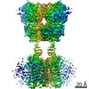
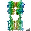
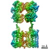
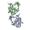
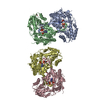
| Entry | Database: PDB / ID: 6w2x | |||||||||
|---|---|---|---|---|---|---|---|---|---|---|
| Title | CryoEM Structure of Inactive GABAB Heterodimer | |||||||||
 Components Components | (Gamma-aminobutyric acid type B receptor subunit ...) x 2 | |||||||||
 Keywords Keywords | SIGNALING PROTEIN / Inhibitor / Heterodimer / GPCR | |||||||||
| Function / homology |  Function and homology information Function and homology informationnegative regulation of gamma-aminobutyric acid secretion / GABA B receptor activation / G protein-coupled GABA receptor complex / : / neuron-glial cell signaling / G protein-coupled receptor heterodimeric complex / G protein-coupled GABA receptor activity / : / negative regulation of epinephrine secretion / negative regulation of dopamine secretion ...negative regulation of gamma-aminobutyric acid secretion / GABA B receptor activation / G protein-coupled GABA receptor complex / : / neuron-glial cell signaling / G protein-coupled receptor heterodimeric complex / G protein-coupled GABA receptor activity / : / negative regulation of epinephrine secretion / negative regulation of dopamine secretion / positive regulation of growth hormone secretion / extracellular matrix protein binding / GABA receptor complex / negative regulation of adenylate cyclase activity / negative regulation of synaptic transmission / Class C/3 (Metabotropic glutamate/pheromone receptors) / gamma-aminobutyric acid signaling pathway / synaptic transmission, GABAergic / positive regulation of glutamate secretion / axolemma / dendritic shaft / response to nicotine / Schaffer collateral - CA1 synapse / GABA-ergic synapse / mitochondrial membrane / osteoblast differentiation / adenylate cyclase-inhibiting G protein-coupled receptor signaling pathway / Activation of G protein gated Potassium channels / Inhibition of voltage gated Ca2+ channels via Gbeta/gamma subunits / transmembrane signaling receptor activity / synaptic vesicle / presynaptic membrane / chemical synaptic transmission / G alpha (i) signalling events / dendritic spine / response to ethanol / postsynaptic membrane / neuron projection / G protein-coupled receptor signaling pathway / protein heterodimerization activity / negative regulation of cell population proliferation / neuronal cell body / endoplasmic reticulum membrane / glutamatergic synapse / : / plasma membrane / cytoplasm Similarity search - Function | |||||||||
| Biological species |  Homo sapiens (human) Homo sapiens (human) | |||||||||
| Method | ELECTRON MICROSCOPY / single particle reconstruction / cryo EM / Resolution: 3.6 Å | |||||||||
 Authors Authors | Papasergi-Scott, M.M. / Robertson, M.J. / Skiniotis, G. | |||||||||
| Funding support |  United States, 1items United States, 1items
| |||||||||
 Citation Citation | Journal: Nature / Year: 2020 Title: Structures of metabotropic GABA receptor. Authors: Makaía M Papasergi-Scott / Michael J Robertson / Alpay B Seven / Ouliana Panova / Jesper M Mathiesen / Georgios Skiniotis /  Abstract: Stimulation of the metabotropic GABA receptor by γ-aminobutyric acid (GABA) results in prolonged inhibition of neurotransmission, which is central to brain physiology. GABA belongs to family C of ...Stimulation of the metabotropic GABA receptor by γ-aminobutyric acid (GABA) results in prolonged inhibition of neurotransmission, which is central to brain physiology. GABA belongs to family C of the G-protein-coupled receptors, which operate as dimers to transform synaptic neurotransmitter signals into a cellular response through the binding and activation of heterotrimeric G proteins. However, GABA is unique in its function as an obligate heterodimer in which agonist binding and G-protein activation take place on distinct subunits. Here we present cryo-electron microscopy structures of heterodimeric and homodimeric full-length GABA receptors. Complemented by cellular signalling assays and atomistic simulations, these structures reveal that extracellular loop 2 (ECL2) of GABA has an essential role in relaying structural transitions by ordering the linker that connects the extracellular ligand-binding domain to the transmembrane region. Furthermore, the ECL2 of each of the subunits of GABA caps and interacts with the hydrophilic head of a phospholipid that occupies the extracellular half of the transmembrane domain, thereby providing a potentially crucial link between ligand binding and the receptor core that engages G proteins. These results provide a starting framework through which to decipher the mechanistic modes of signal transduction mediated by GABA dimers, and have important implications for rational drug design that targets these receptors. | |||||||||
| History |
|
- Structure visualization
| Movie |
Movie viewer |
|---|---|
| Structure viewer | Molecule: MolmilJmol/JSmol |
- Downloads & links
-Download
| PDBx/mmCIF format | 6w2x.cif.gz | 254.3 KB | Display | PDBx/mmCIF format |
|---|---|---|---|---|
| PDB format | pdb6w2x.ent.gz | 186 KB | Display | PDB format |
| PDBx/mmJSON format | 6w2x.json.gz | Tree view | PDBx/mmJSON format | |
| Others |  Other downloads Other downloads |
-Validation report
| Arichive directory | https://data.pdbj.org/pub/pdb/validation_reports/w2/6w2xftp://data.pdbj.org/pub/pdb/validation_reports/w2/6w2x | HTTPS FTP |
|---|
-Related structure data
| Related structure data | 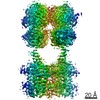 21533MC 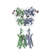 6w2yC M: map data used to model this data C: citing same article ( |
|---|---|
| Similar structure data |
-Links
 PDBj
PDBj


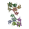
- Assembly
| Deposited unit | 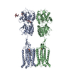
|
|---|---|
| 1 |
|
-Components
-Gamma-aminobutyric acid type B receptor subunit ... , 2 types, 2 molecules AB
| #1: Protein | Mass: 94173.570 Da / Num. of mol.: 1 / Fragment: UNP residues 30-844 Source method: isolated from a genetically manipulated source Source: (gene. exp.) Homo sapiens (human) / Gene: GABBR1, GPRC3A / Production host:   Spodoptera frugiperda (fall armyworm) / References: UniProt: Q9UBS5 Spodoptera frugiperda (fall armyworm) / References: UniProt: Q9UBS5 |
|---|---|
| #2: Protein | Mass: 102799.164 Da / Num. of mol.: 1 / Fragment: UNP residues 42-941 Source method: isolated from a genetically manipulated source Source: (gene. exp.) Homo sapiens (human) / Gene: GABBR2, GPR51, GPRC3B / Production host: Spodoptera frugiperda (fall armyworm) / References: UniProt: O75899 |
-Sugars , 2 types, 4 molecules 
| #3: Polysaccharide | Source method: isolated from a genetically manipulated source #4: Sugar |  Type: D-saccharide, beta linking / Mass: 221.208 Da / Num. of mol.: 2 Type: D-saccharide, beta linking / Mass: 221.208 Da / Num. of mol.: 2Source method: isolated from a genetically manipulated source Formula: C8H15NO6 |
|---|
-Non-polymers , 3 types, 4 molecules 
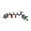

| #5: Chemical |  Mass: 746.050 Da / Num. of mol.: 2 / Source method: obtained synthetically / Formula: C41H80NO8P Mass: 746.050 Da / Num. of mol.: 2 / Source method: obtained synthetically / Formula: C41H80NO8P#6: Chemical | ChemComp-SGG / [( |  Mass: 402.252 Da / Num. of mol.: 1 / Source method: obtained synthetically / Formula: C18H22Cl2NO3P Mass: 402.252 Da / Num. of mol.: 1 / Source method: obtained synthetically / Formula: C18H22Cl2NO3P#7: Chemical | ChemComp-MG / |  Mass: 24.305 Da / Num. of mol.: 1 / Source method: obtained synthetically / Formula: Mg Mass: 24.305 Da / Num. of mol.: 1 / Source method: obtained synthetically / Formula: Mg |
|---|
-Details
| Has ligand of interest | N |
|---|---|
| Has protein modification | Y |
-Experimental details
-Experiment
| Experiment | Method: ELECTRON MICROSCOPY |
|---|---|
| EM experiment | Aggregation state: PARTICLE / 3D reconstruction method: single particle reconstruction |
- Sample preparation
| Component | Name: GABAB Heterodimer / Type: COMPLEX / Entity ID: #1-#2 / Source: RECOMBINANT | |||||||||||||||||||||||||
|---|---|---|---|---|---|---|---|---|---|---|---|---|---|---|---|---|---|---|---|---|---|---|---|---|---|---|
| Molecular weight | Experimental value: NO | |||||||||||||||||||||||||
| Source (natural) | Organism: Homo sapiens (human) | |||||||||||||||||||||||||
| Source (recombinant) | Organism: Spodoptera frugiperda (fall armyworm) | |||||||||||||||||||||||||
| Buffer solution | pH: 7.5 | |||||||||||||||||||||||||
| Buffer component |
| |||||||||||||||||||||||||
| Specimen | Embedding applied: NO / Shadowing applied: NO / Staining applied: NO / Vitrification applied: YES | |||||||||||||||||||||||||
| Specimen support | Grid material: GOLD / Grid type: Quantifoil R1.2/1.3 | |||||||||||||||||||||||||
| Vitrification | Instrument: FEI VITROBOT MARK IV / Cryogen name: ETHANE / Humidity: 100 % / Chamber temperature: 291 K |
- Electron microscopy imaging
| Experimental equipment |  Model: Titan Krios / Image courtesy: FEI Company |
|---|---|
| Microscopy | Model: TFS KRIOS |
| Electron gun | Electron source:  FIELD EMISSION GUN / Accelerating voltage: 300 kV / Illumination mode: OTHER FIELD EMISSION GUN / Accelerating voltage: 300 kV / Illumination mode: OTHER |
| Electron lens | Mode: BRIGHT FIELD |
| Image recording | Average exposure time: 3.5 sec. / Electron dose: 60.5 e/Å2 / Film or detector model: GATAN K3 (6k x 4k) |
- Processing
| EM software |
| ||||||||||||||||||||||||||||||||||||
|---|---|---|---|---|---|---|---|---|---|---|---|---|---|---|---|---|---|---|---|---|---|---|---|---|---|---|---|---|---|---|---|---|---|---|---|---|---|
| CTF correction | Type: NONE | ||||||||||||||||||||||||||||||||||||
| Particle selection | Num. of particles selected: 2601040 | ||||||||||||||||||||||||||||||||||||
| Symmetry | Point symmetry: C1 (asymmetric) | ||||||||||||||||||||||||||||||||||||
| 3D reconstruction | Resolution: 3.6 Å / Resolution method: FSC 0.143 CUT-OFF / Num. of particles: 286140 / Symmetry type: POINT |
