 Movie
Movie Controller

[English] 日本語
 Yorodumi
- PDB-6vw2: Cryo-EM structure of human islet amyloid polypeptide (hIAPP, or a... -
+ Open data

- Basic information
| Entry | Database: PDB / ID: 6vw2 | ||||||
|---|---|---|---|---|---|---|---|
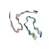
| Title | Cryo-EM structure of human islet amyloid polypeptide (hIAPP, or amylin) fibrils | ||||||
 Components Components | Islet amyloid polypeptide (SUMO-tagged) | ||||||
 Keywords Keywords | PROTEIN FIBRIL / hIAPP / type II diabetes / amyloid | ||||||
| Function / homology |  Function and homology information Function and homology informationSUMO is conjugated to E1 (UBA2:SAE1) / SUMOylation of nuclear envelope proteins / SUMO is transferred from E1 to E2 (UBE2I, UBC9) / SUMO is proteolytically processed / SUMOylation of transcription factors / Postmitotic nuclear pore complex (NPC) reformation / amylin receptor 3 signaling pathway / amylin receptor 2 signaling pathway / SUMOylation of transcription cofactors / septin ring ...SUMO is conjugated to E1 (UBA2:SAE1) / SUMOylation of nuclear envelope proteins / SUMO is transferred from E1 to E2 (UBE2I, UBC9) / SUMO is proteolytically processed / SUMOylation of transcription factors / Postmitotic nuclear pore complex (NPC) reformation / amylin receptor 3 signaling pathway / amylin receptor 2 signaling pathway / SUMOylation of transcription cofactors / septin ring / amylin receptor 1 signaling pathway / SUMOylation of DNA damage response and repair proteins / amylin receptor signaling pathway / Calcitonin-like ligand receptors / Transcriptional and post-translational regulation of MITF-M expression and activity / SUMOylation of DNA replication proteins / SUMOylation of SUMOylation proteins / Recruitment and ATM-mediated phosphorylation of repair and signaling proteins at DNA double strand breaks / negative regulation of amyloid fibril formation / SUMOylation of RNA binding proteins / negative regulation of bone resorption / SUMOylation of chromatin organization proteins / eating behavior / negative regulation of osteoclast differentiation / Regulation of gene expression in beta cells / positive regulation of cAMP/PKA signal transduction / ubiquitin-like protein ligase binding / protein sumoylation / bone resorption / negative regulation of protein-containing complex assembly / positive regulation of calcium-mediated signaling / osteoclast differentiation / sensory perception of pain / condensed nuclear chromosome / hormone activity / protein tag activity / cell-cell signaling / amyloid-beta binding / G alpha (s) signalling events / positive regulation of MAPK cascade / positive regulation of apoptotic process / Amyloid fiber formation / receptor ligand activity / signaling receptor binding / neuronal cell body / apoptotic process / lipid binding / signal transduction / : / extracellular region / identical protein binding / nucleus Similarity search - Function | ||||||
| Biological species |   Homo sapiens (human) Homo sapiens (human) | ||||||
| Method | ELECTRON MICROSCOPY / helical reconstruction / cryo EM / Resolution: 3.4 Å | ||||||
 Authors Authors | Cao, Q. / Boyer, D.R. / Sawaya, M.R. / Eisenberg, D.S. | ||||||
| Funding support |  United States, 1items United States, 1items
| ||||||
 Citation Citation | Journal: Nat Struct Mol Biol / Year: 2020 Title: Cryo-EM structure and inhibitor design of human IAPP (amylin) fibrils. Authors: Qin Cao / David R Boyer / Michael R Sawaya / Peng Ge / David S Eisenberg / Abstract: Human islet amyloid polypeptide (hIAPP) functions as a glucose-regulating hormone but deposits as amyloid fibrils in more than 90% of patients with type II diabetes (T2D). Here we report the cryo-EM ...Human islet amyloid polypeptide (hIAPP) functions as a glucose-regulating hormone but deposits as amyloid fibrils in more than 90% of patients with type II diabetes (T2D). Here we report the cryo-EM structure of recombinant full-length hIAPP fibrils. The fibril is composed of two symmetrically related protofilaments with ordered residues 14-37. Our hIAPP fibril structure (i) supports the previous hypothesis that residues 20-29 constitute the core of the hIAPP amyloid; (ii) suggests a molecular mechanism for the action of the hIAPP hereditary mutation S20G; (iii) explains why the six residue substitutions in rodent IAPP prevent aggregation; and (iv) suggests regions responsible for the observed hIAPP cross-seeding with β-amyloid. Furthermore, we performed structure-based inhibitor design to generate potential hIAPP aggregation inhibitors. Four of the designed peptides delay hIAPP aggregation in vitro, providing a starting point for the development of T2D therapeutics and proof of concept that the capping strategy can be used on full-length cryo-EM fibril structures. | ||||||
| History |
|
- Structure visualization
| Movie |
Movie viewer |
|---|---|
| Structure viewer | Molecule: MolmilJmol/JSmol |
- Downloads & links
-Download
| PDBx/mmCIF format | 6vw2.cif.gz | 67.9 KB | Display | PDBx/mmCIF format |
|---|---|---|---|---|
| PDB format | pdb6vw2.ent.gz | 41.1 KB | Display | PDB format |
| PDBx/mmJSON format | 6vw2.json.gz | Tree view | PDBx/mmJSON format | |
| Others |  Other downloads Other downloads |
-Validation report
| Arichive directory | https://data.pdbj.org/pub/pdb/validation_reports/vw/6vw2ftp://data.pdbj.org/pub/pdb/validation_reports/vw/6vw2 | HTTPS FTP |
|---|
-Related structure data
| Related structure data |  21410MC M: map data used to model this data C: citing same article ( |
|---|---|
| Similar structure data | |
| EM raw data | EMPIAR-10871 (Title: Cryo electron microscopy of recombinant, un-seeded hIAPP fibrils Data size: 1.9 TB Data #1: Unaligned K2 movies of unseeded, recombinant hIAPP amyloid fibrils [micrographs - multiframe]) |


-Links
 PDBj
PDBj


- Assembly
| Deposited unit | 
|
|---|---|
| 1 |
|
-Components
| #1: Protein | Mass: 17274.273 Da / Num. of mol.: 10 Source method: isolated from a genetically manipulated source Source: (gene. exp.) Homo sapiens (human)Strain: ATCC 204508 / S288c / Gene: SMT3, YDR510W, D9719.15, IAPP / Production host:  |
|---|
-Experimental details
-Experiment
| Experiment | Method: ELECTRON MICROSCOPY |
|---|---|
| EM experiment | Aggregation state: FILAMENT / 3D reconstruction method: helical reconstruction |
- Sample preparation
| Component | Name: hIAPP fibril / Type: ORGANELLE OR CELLULAR COMPONENT / Entity ID: all / Source: RECOMBINANT |
|---|---|
| Source (natural) | Organism: Homo sapiens (human) |
| Source (recombinant) | Organism: |
| Buffer solution | pH: 7.4 |
| Specimen | Embedding applied: NO / Shadowing applied: NO / Staining applied: NO / Vitrification applied: YES |
| Vitrification | Cryogen name: ETHANE |
- Electron microscopy imaging
| Experimental equipment |  Model: Titan Krios / Image courtesy: FEI Company |
|---|---|
| Microscopy | Model: FEI TITAN KRIOS |
| Electron gun | Electron source:  FIELD EMISSION GUN / Accelerating voltage: 300 kV / Illumination mode: FLOOD BEAM FIELD EMISSION GUN / Accelerating voltage: 300 kV / Illumination mode: FLOOD BEAM |
| Electron lens | Mode: BRIGHT FIELD / Nominal defocus max: 2000 nm / Nominal defocus min: 2000 nm / Cs: 2.7 mm / C2 aperture diameter: 50 µm |
| Image recording | Average exposure time: 8 sec. / Electron dose: 44 e/Å2 / Detector mode: SUPER-RESOLUTION / Film or detector model: GATAN K2 SUMMIT (4k x 4k) |
- Processing
| EM software |
| ||||||||||||||||||||||||||||||||||||||||
|---|---|---|---|---|---|---|---|---|---|---|---|---|---|---|---|---|---|---|---|---|---|---|---|---|---|---|---|---|---|---|---|---|---|---|---|---|---|---|---|---|---|
| CTF correction | Type: PHASE FLIPPING AND AMPLITUDE CORRECTION | ||||||||||||||||||||||||||||||||||||||||
| Helical symmerty | Angular rotation/subunit: 179.42 ° / Axial rise/subunit: 2.406 Å / Axial symmetry: C1 | ||||||||||||||||||||||||||||||||||||||||
| 3D reconstruction | Resolution: 3.4 Å / Resolution method: FSC 0.143 CUT-OFF / Num. of particles: 25767 / Symmetry type: HELICAL |
