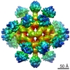
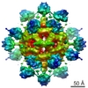
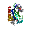
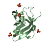
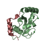
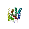
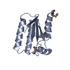
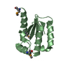
Journal: Science / Year: 2008 Title: Molecular architecture of the "stressosome," a signal integration and transduction hub. Authors: Jon Marles-Wright / Tim Grant / Olivier Delumeau / Gijs van Duinen / Susan J Firbank / Peter J Lewis / James W Murray / Joseph A Newman / Maureen B Quin / Paul R Race / Alexis Rohou / Willem ...Authors: Jon Marles-Wright / Tim Grant / Olivier Delumeau / Gijs van Duinen / Susan J Firbank / Peter J Lewis / James W Murray / Joseph A Newman / Maureen B Quin / Paul R Race / Alexis Rohou / Willem Tichelaar / Marin van Heel / Richard J Lewis / Abstract: A commonly used strategy by microorganisms to survive multiple stresses involves a signal transduction cascade that increases the expression of stress-responsive genes. Stress signals can be ...A commonly used strategy by microorganisms to survive multiple stresses involves a signal transduction cascade that increases the expression of stress-responsive genes. Stress signals can be integrated by a multiprotein signaling hub that responds to various signals to effect a single outcome. We obtained a medium-resolution cryo-electron microscopy reconstruction of the 1.8-megadalton "stressosome" from Bacillus subtilis. Fitting known crystal structures of components into this reconstruction gave a pseudoatomic structure, which had a virus capsid-like core with sensory extensions. We suggest that the different sensory extensions respond to different signals, whereas the conserved domains in the core integrate the varied signals. The architecture of the stressosome provides the potential for cooperativity, suggesting that the response could be tuned dependent on the magnitude of chemophysical insult.
Protocol: SINGLE WAVELENGTH / Monochromatic (M) / Laue (L): M / Scattering type: x-ray
Radiation wavelength
Wavelength: 0.9699 Å / Relative weight: 1
Reflection
Resolution: 2.3→19.1 Å / Num. obs: 12304 / % possible obs: 97.2 % / Observed criterion σ(I): 1.5 / Redundancy: 3.4 % / Rmerge(I) obs: 0.08 / Net I/σ(I): 11.1
Reflection shell
Resolution: 2.3→2.42 Å / Redundancy: 3.5 % / Rmerge(I) obs: 0.54 / Mean I/σ(I) obs: 2.1 / % possible all: 98.8
-
Processing
Software
Name
Version
Classification
REFMAC
5.4.0077
refinement
MOSFLM
datareduction
SCALA
datascaling
SHELX
phasing
Refinement
Method to determine structure: SAD Starting model: NONE Resolution: 2.3→19.06 Å / Cor.coef. Fo:Fc: 0.935 / Cor.coef. Fo:Fc free: 0.921 / SU B: 21.491 / SU ML: 0.22 / TLS residual ADP flag: LIKELY RESIDUAL / Cross valid method: THROUGHOUT / ESU R: 0.341 / ESU R Free: 0.261 / Stereochemistry target values: MAXIMUM LIKELIHOOD Details: HYDROGENS HAVE BEEN ADDED IN THE RIDING POSITIONS. U VALUES RESIDUAL ONLY
Rfactor
Num. reflection
% reflection
Selection details
Rfree
0.28
593
4.8 %
RANDOM
Rwork
0.226
-
-
-
obs
0.228
11710
96.3 %
-
Solvent computation
Ion probe radii: 0.8 Å / Shrinkage radii: 0.8 Å / VDW probe radii: 1.2 Å / Solvent model: MASK
 Movie
Movie Controller

 Yorodumi
 Open data
 Basic information
 Components
 Keywords
 Function and homology information
 MOORELLA THERMOACETICA (bacteria)
 X-RAY DIFFRACTION /
  Authors
 Citation
 Structure visualization
 Downloads & links
 Other downloads
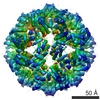


 PDBj
PDBj
 Assembly
 Mass: 18.015 Da / Num. of mol.: 42 / Source method: isolated from a natural source / Formula: H2O
 Sample preparation
 Processing