 Movie
Movie Controller


+ Open data

- Basic information
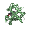
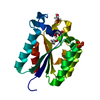
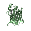
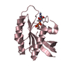
| Entry | Database: PDB / ID: 5yir | ||||||
|---|---|---|---|---|---|---|---|
| Title | Crystal Structure of AnkB LIR/GABARAP complex | ||||||
 Components Components |
| ||||||
 Keywords Keywords | PROTEIN BINDING / Autophagy | ||||||
| Function / homology |  Function and homology information Function and homology informationprotein localization to T-tubule / atrial cardiac muscle cell to AV node cell communication / SA node cell to atrial cardiac muscle cell communication / TBC/RABGAPs / Macroautophagy / protein localization to M-band / T-tubule organization / SA node cell action potential / membrane depolarization during SA node cell action potential / paranodal junction assembly ...protein localization to T-tubule / atrial cardiac muscle cell to AV node cell communication / SA node cell to atrial cardiac muscle cell communication / TBC/RABGAPs / Macroautophagy / protein localization to M-band / T-tubule organization / SA node cell action potential / membrane depolarization during SA node cell action potential / paranodal junction assembly / positive regulation of protein K48-linked ubiquitination / potassium channel activator activity / positive regulation of potassium ion import across plasma membrane / regulation of atrial cardiac muscle cell action potential / regulation of Rac protein signal transduction / channel activator activity / phosphorylation-dependent protein binding / sarcoplasmic reticulum calcium ion transport / regulation of SA node cell action potential / atrial cardiac muscle cell action potential / protein localization to endoplasmic reticulum / A band / atrial septum development / cytoskeletal adaptor activity / costamere / ventricular cardiac muscle cell action potential / GABA receptor binding / phosphatidylethanolamine binding / regulation of ventricular cardiac muscle cell membrane repolarization / positive regulation of calcium ion transport / response to methylmercury / regulation of release of sequestered calcium ion into cytosol / regulation of cardiac muscle cell contraction / protein localization to cell surface / M band / postsynaptic specialization, intracellular component / cellular response to nitrogen starvation / microtubule associated complex / regulation of cardiac muscle contraction by calcium ion signaling / Interaction between L1 and Ankyrins / spectrin binding / regulation of neurotransmitter receptor localization to postsynaptic specialization membrane / smooth endoplasmic reticulum / regulation of calcium ion transport / regulation of heart rate by cardiac conduction / autophagosome membrane / intercalated disc / extrinsic apoptotic signaling pathway via death domain receptors / autophagosome maturation / axoneme / regulation of cardiac muscle contraction / autophagosome assembly / beta-tubulin binding / mitophagy / COPI-mediated anterograde transport / regulation of cardiac muscle contraction by regulation of the release of sequestered calcium ion / T-tubule / regulation of heart rate / autophagosome / protein localization to plasma membrane / regulation of protein stability / sarcolemma / recycling endosome / structural constituent of cytoskeleton / microtubule cytoskeleton organization / GABA-ergic synapse / Z disc / endocytosis / intracellular calcium ion homeostasis / intracellular protein localization / positive regulation of proteasomal ubiquitin-dependent protein catabolic process / protein transport / actin cytoskeleton / ATPase binding / cell body / cytoplasmic vesicle / sperm midpiece / microtubule binding / basolateral plasma membrane / cytoskeleton / microtubule / transmembrane transporter binding / protein-macromolecule adaptor activity / early endosome / postsynaptic membrane / lysosome / apical plasma membrane / neuron projection / protein stabilization / Golgi membrane / ubiquitin protein ligase binding / positive regulation of gene expression / protein kinase binding / perinuclear region of cytoplasm / enzyme binding / Golgi apparatus / mitochondrion / plasma membrane / cytosol Similarity search - Function | ||||||
| Biological species |   Homo sapiens (human) Homo sapiens (human) | ||||||
| Method |  X-RAY DIFFRACTION / SYNCHROTRON / MOLECULAR REPLACEMENT / Resolution: 2.75 Å X-RAY DIFFRACTION / SYNCHROTRON / MOLECULAR REPLACEMENT / Resolution: 2.75 Å | ||||||
 Authors Authors | Li, J. / Zhu, R. / Chen, K. / Zheng, H. / Yuan, C. / Zhang, H. / Wang, C. / Zhang, M. | ||||||
| Funding support |  Hong Kong, 1items Hong Kong, 1items
| ||||||
 Citation Citation | Journal: Nat. Chem. Biol. / Year: 2018 Title: Potent and specific Atg8-targeting autophagy inhibitory peptides from giant ankyrins. Authors: Li, J. / Zhu, R. / Chen, K. / Zheng, H. / Zhao, H. / Yuan, C. / Zhang, H. / Wang, C. / Zhang, M. | ||||||
| History |
|
- Structure visualization
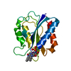
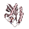
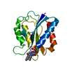
| Structure viewer | Molecule: MolmilJmol/JSmol |
|---|
- Downloads & links
-Download
| PDBx/mmCIF format | 5yir.cif.gz | 99.6 KB | Display | PDBx/mmCIF format |
|---|---|---|---|---|
| PDB format | pdb5yir.ent.gz | 73.2 KB | Display | PDB format |
| PDBx/mmJSON format | 5yir.json.gz | Tree view | PDBx/mmJSON format | |
| Others |  Other downloads Other downloads |
-Validation report
| Arichive directory | https://data.pdbj.org/pub/pdb/validation_reports/yi/5yirftp://data.pdbj.org/pub/pdb/validation_reports/yi/5yir | HTTPS FTP |
|---|
-Related structure data
| Related structure data |  5yipC 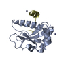 5yiqC  5yisC  1kjtS S: Starting model for refinement C: citing same article ( |
|---|---|
| Similar structure data |


-Links
 PDBj
PDBj


- Assembly
| Deposited unit | 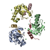
| |||||||||||||||||||||||||||||||||||||||||||||||||||||||||||||||||||||||||||||||||||||||||||
|---|---|---|---|---|---|---|---|---|---|---|---|---|---|---|---|---|---|---|---|---|---|---|---|---|---|---|---|---|---|---|---|---|---|---|---|---|---|---|---|---|---|---|---|---|---|---|---|---|---|---|---|---|---|---|---|---|---|---|---|---|---|---|---|---|---|---|---|---|---|---|---|---|---|---|---|---|---|---|---|---|---|---|---|---|---|---|---|---|---|---|---|---|
| 1 | 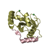
| |||||||||||||||||||||||||||||||||||||||||||||||||||||||||||||||||||||||||||||||||||||||||||
| 2 | 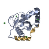
| |||||||||||||||||||||||||||||||||||||||||||||||||||||||||||||||||||||||||||||||||||||||||||
| 3 | 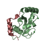
| |||||||||||||||||||||||||||||||||||||||||||||||||||||||||||||||||||||||||||||||||||||||||||
| Unit cell |
| |||||||||||||||||||||||||||||||||||||||||||||||||||||||||||||||||||||||||||||||||||||||||||
| Noncrystallographic symmetry (NCS) | NCS domain:
NCS domain segments:
|