 Movie
Movie Controller

[English] 日本語
 Yorodumi
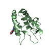
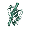
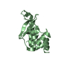
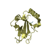
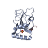
- PDB-1kjt: Crystal Structure of the GABA(A) Receptor Associated Protein, GABARAP -
+ Open data

- Basic information
| Entry | Database: PDB / ID: 1kjt | ||||||
|---|---|---|---|---|---|---|---|
| Title | Crystal Structure of the GABA(A) Receptor Associated Protein, GABARAP | ||||||
 Components Components | GABARAP | ||||||
 Keywords Keywords | TRANSPORT PROTEIN / ubiquitin-like fold / N-terminal alpha helical region | ||||||
| Function / homology |  Function and homology information Function and homology informationTBC/RABGAPs / Macroautophagy / positive regulation of protein K48-linked ubiquitination / regulation of Rac protein signal transduction / GABA receptor binding / phosphatidylethanolamine binding / postsynaptic specialization, intracellular component / cellular response to nitrogen starvation / microtubule associated complex / regulation of neurotransmitter receptor localization to postsynaptic specialization membrane ...TBC/RABGAPs / Macroautophagy / positive regulation of protein K48-linked ubiquitination / regulation of Rac protein signal transduction / GABA receptor binding / phosphatidylethanolamine binding / postsynaptic specialization, intracellular component / cellular response to nitrogen starvation / microtubule associated complex / regulation of neurotransmitter receptor localization to postsynaptic specialization membrane / smooth endoplasmic reticulum / autophagosome membrane / extrinsic apoptotic signaling pathway via death domain receptors / autophagosome maturation / axoneme / autophagosome assembly / beta-tubulin binding / mitophagy / autophagosome / microtubule cytoskeleton organization / GABA-ergic synapse / protein transport / positive regulation of proteasomal ubiquitin-dependent protein catabolic process / actin cytoskeleton / cell body / cytoplasmic vesicle / sperm midpiece / microtubule binding / microtubule / lysosome / Golgi membrane / ubiquitin protein ligase binding / perinuclear region of cytoplasm / Golgi apparatus / plasma membrane Similarity search - Function | ||||||
| Biological species |  | ||||||
| Method |  X-RAY DIFFRACTION / SYNCHROTRON / MOLECULAR REPLACEMENT / Resolution: 2 Å X-RAY DIFFRACTION / SYNCHROTRON / MOLECULAR REPLACEMENT / Resolution: 2 Å | ||||||
 Authors Authors | Bavro, V.N. / Sola, M. / Bracher, A. / Kneussel, M. / Betz, H. / Weissenhorn, W. | ||||||
 Citation Citation | Journal: EMBO Rep. / Year: 2002 Title: Crystal structure of the GABA(A)-receptor-associated protein, GABARAP. Authors: Bavro, V.N. / Sola, M. / Bracher, A. / Kneussel, M. / Betz, H. / Weissenhorn, W. | ||||||
| History |
|
- Structure visualization
| Structure viewer | Molecule: MolmilJmol/JSmol |
|---|
- Downloads & links
-Download
| PDBx/mmCIF format | 1kjt.cif.gz | 37.3 KB | Display | PDBx/mmCIF format |
|---|---|---|---|---|
| PDB format | pdb1kjt.ent.gz | 25.6 KB | Display | PDB format |
| PDBx/mmJSON format | 1kjt.json.gz | Tree view | PDBx/mmJSON format | |
| Others |  Other downloads Other downloads |
-Validation report
| Arichive directory | https://data.pdbj.org/pub/pdb/validation_reports/kj/1kjtftp://data.pdbj.org/pub/pdb/validation_reports/kj/1kjt | HTTPS FTP |
|---|
-Related structure data
| Similar structure data |
|---|

-Links
 PDBj
PDBj
- Assembly
| Deposited unit | 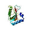
| ||||||||
|---|---|---|---|---|---|---|---|---|---|
| 1 |
| ||||||||
| Unit cell |
|
-Components
| #1: Protein | Mass: 14100.201 Da / Num. of mol.: 1 Source method: isolated from a genetically manipulated source Source: (gene. exp.)  | ||||
|---|---|---|---|---|---|
| #2: Chemical |   Mass: 58.693 Da / Num. of mol.: 2 / Source method: obtained synthetically / Formula: Ni Mass: 58.693 Da / Num. of mol.: 2 / Source method: obtained synthetically / Formula: Ni#3: Chemical | ChemComp-NA / |   Mass: 22.990 Da / Num. of mol.: 1 / Source method: obtained synthetically / Formula: Na Mass: 22.990 Da / Num. of mol.: 1 / Source method: obtained synthetically / Formula: Na#4: Water | ChemComp-HOH / |  Mass: 18.015 Da / Num. of mol.: 52 / Source method: isolated from a natural source / Formula: H2O Mass: 18.015 Da / Num. of mol.: 52 / Source method: isolated from a natural source / Formula: H2O |
-Experimental details
-Experiment
| Experiment | Method: X-RAY DIFFRACTION / Number of used crystals: 1 |
|---|
- Sample preparation
| Crystal | Density Matthews: 1.92 Å3/Da / Density % sol: 35.88 % | |||||||||||||||||||||||||||||||||||||||||||||||||
|---|---|---|---|---|---|---|---|---|---|---|---|---|---|---|---|---|---|---|---|---|---|---|---|---|---|---|---|---|---|---|---|---|---|---|---|---|---|---|---|---|---|---|---|---|---|---|---|---|---|---|
| Crystal grow | Temperature: 293 K / Method: vapor diffusion, hanging drop / pH: 8.5 Details: PEG 2000MME, Tris HCL, nickel chloride, sodium chloride, pH 8.5, VAPOR DIFFUSION, HANGING DROP, temperature 293K | |||||||||||||||||||||||||||||||||||||||||||||||||
| Crystal grow | *PLUS pH: 7.2 / Method: vapor diffusion | |||||||||||||||||||||||||||||||||||||||||||||||||
| Components of the solutions | *PLUS
|
-Data collection
| Diffraction | Mean temperature: 100 K |
|---|---|
| Diffraction source | Source: SYNCHROTRON / Site: ESRF  / Beamline: ID14-2 / Wavelength: 0.9333 Å / Beamline: ID14-2 / Wavelength: 0.9333 Å |
| Detector | Type: ADSC QUANTUM 4 / Detector: CCD / Date: Sep 20, 2001 |
| Radiation | Monochromator: DIAMOND / Protocol: SINGLE WAVELENGTH / Monochromatic (M) / Laue (L): M / Scattering type: x-ray |
| Radiation wavelength | Wavelength: 0.9333 Å / Relative weight: 1 |
| Reflection | Resolution: 2→30 Å / Num. all: 7790 / Num. obs: 7736 |
| Reflection shell | Resolution: 2→2.07 Å |
| Reflection | *PLUS Num. obs: 7790 / % possible obs: 99.4 % / Redundancy: 3.8 % / Num. measured all: 56243 / Rmerge(I) obs: 0.05 |
| Reflection shell | *PLUS Highest resolution: 2 Å / % possible obs: 95.5 % / Redundancy: 3.3 % / Rmerge(I) obs: 0.439 / Mean I/σ(I) obs: 2.11 |
- Processing
| Software |
| ||||||||||||||||
|---|---|---|---|---|---|---|---|---|---|---|---|---|---|---|---|---|---|
| Refinement | Method to determine structure: MOLECULAR REPLACEMENT / Resolution: 2→30 Å / σ(F): 0 / Stereochemistry target values: Engh & Huber
| ||||||||||||||||
| Refinement step | Cycle: LAST / Resolution: 2→30 Å
| ||||||||||||||||
| Refinement | *PLUS Lowest resolution: 30 Å / Rfactor obs: 0.235 / Rfactor Rfree: 0.261 / Rfactor Rwork: 0.235 | ||||||||||||||||
| Solvent computation | *PLUS | ||||||||||||||||
| Displacement parameters | *PLUS | ||||||||||||||||
| Refine LS restraints | *PLUS
|
