 Movie
Movie Controller


+ Open data

- Basic information
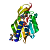
| Entry | Database: PDB / ID: 1gnu | ||||||
|---|---|---|---|---|---|---|---|
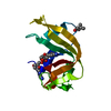
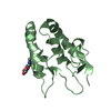
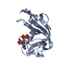
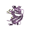
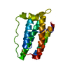
| Title | GABA(A) receptor associated protein GABARAP | ||||||
 Components Components | GABARAP | ||||||
 Keywords Keywords | TRANSPORT / UBIQUITIN-LIKE / RECEPTOR | ||||||
| Function / homology |  Function and homology information Function and homology informationpositive regulation of protein K48-linked ubiquitination / regulation of Rac protein signal transduction / GABA receptor binding / phosphatidylethanolamine binding / TBC/RABGAPs / cellular response to nitrogen starvation / microtubule associated complex / Macroautophagy / regulation of neurotransmitter receptor localization to postsynaptic specialization membrane / smooth endoplasmic reticulum ...positive regulation of protein K48-linked ubiquitination / regulation of Rac protein signal transduction / GABA receptor binding / phosphatidylethanolamine binding / TBC/RABGAPs / cellular response to nitrogen starvation / microtubule associated complex / Macroautophagy / regulation of neurotransmitter receptor localization to postsynaptic specialization membrane / smooth endoplasmic reticulum / autophagosome membrane / extrinsic apoptotic signaling pathway via death domain receptors / autophagosome maturation / axoneme / autophagosome assembly / beta-tubulin binding / mitophagy / protein targeting / autophagosome / microtubule cytoskeleton organization / GABA-ergic synapse / positive regulation of proteasomal ubiquitin-dependent protein catabolic process / protein transport / actin cytoskeleton / cytoplasmic vesicle / sperm midpiece / microtubule binding / microtubule / chemical synaptic transmission / lysosome / Golgi membrane / ubiquitin protein ligase binding / plasma membrane / cytosol Similarity search - Function | ||||||
| Biological species |  HOMO SAPIENS (human) HOMO SAPIENS (human) | ||||||
| Method |  X-RAY DIFFRACTION / SYNCHROTRON / MOLECULAR REPLACEMENT / Resolution: 1.75 Å X-RAY DIFFRACTION / SYNCHROTRON / MOLECULAR REPLACEMENT / Resolution: 1.75 Å | ||||||
 Authors Authors | Knight, D. / Harris, R. / Moss, S. / Driscoll, P.C. / Keep, N.H. | ||||||
 Citation Citation | Journal: J.Biol.Chem. / Year: 2002 Title: The X-Ray Crystal Structure and Putative Ligand-Derived Peptide Binding Properties of Gamma-Aminobutyric Acid Receptor Type a Receptor-Associated Protein Authors: Knight, D. / Harris, R. / Mcalister, M. / Phelan, J. / Geddes, S. / Moss, S. / Driscoll, P.C. / Keep, N.H. | ||||||
| History |
|
- Structure visualization
| Structure viewer | Molecule: MolmilJmol/JSmol |
|---|
- Downloads & links
-Download
| PDBx/mmCIF format | 1gnu.cif.gz | 40.6 KB | Display | PDBx/mmCIF format |
|---|---|---|---|---|
| PDB format | pdb1gnu.ent.gz | 27.7 KB | Display | PDB format |
| PDBx/mmJSON format | 1gnu.json.gz | Tree view | PDBx/mmJSON format | |
| Others |  Other downloads Other downloads |
-Validation report
| Arichive directory | https://data.pdbj.org/pub/pdb/validation_reports/gn/1gnuftp://data.pdbj.org/pub/pdb/validation_reports/gn/1gnu | HTTPS FTP |
|---|
-Related structure data
| Related structure data | 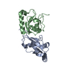 1eo6S S: Starting model for refinement |
|---|---|
| Similar structure data |


-Links
 PDBj
PDBj


- Assembly
| Deposited unit | 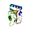
| ||||||||
|---|---|---|---|---|---|---|---|---|---|
| 1 |
| ||||||||
| Unit cell |
|
-Components
| #1: Protein | Mass: 13942.047 Da / Num. of mol.: 1 Source method: isolated from a genetically manipulated source Source: (gene. exp.) HOMO SAPIENS (human) / Tissue: NEURONAL / Production host:  |
|---|---|
| #2: Chemical | ChemComp-NI /   Mass: 58.693 Da / Num. of mol.: 1 / Source method: obtained synthetically / Formula: Ni Mass: 58.693 Da / Num. of mol.: 1 / Source method: obtained synthetically / Formula: Ni |
| #3: Water | ChemComp-HOH /  Mass: 18.015 Da / Num. of mol.: 100 / Source method: isolated from a natural source / Formula: H2O Mass: 18.015 Da / Num. of mol.: 100 / Source method: isolated from a natural source / Formula: H2O |
-Experimental details
-Experiment
| Experiment | Method: X-RAY DIFFRACTION / Number of used crystals: 1 |
|---|
- Sample preparation
| Crystal | Density Matthews: 1.91 Å3/Da / Density % sol: 69 % | |||||||||||||||||||||||||||||||||||||||||||||||||
|---|---|---|---|---|---|---|---|---|---|---|---|---|---|---|---|---|---|---|---|---|---|---|---|---|---|---|---|---|---|---|---|---|---|---|---|---|---|---|---|---|---|---|---|---|---|---|---|---|---|---|
| Crystal grow | pH: 8.5 Details: 18MG/ML GABARAP, 50MM TRIS PH8.5, 0.25M NACL,10%(W/V) PEGMONO, pH 8.50 | |||||||||||||||||||||||||||||||||||||||||||||||||
| Crystal grow | *PLUS Temperature: 22 ℃ / pH: 7 / Method: vapor diffusion, hanging drop | |||||||||||||||||||||||||||||||||||||||||||||||||
| Components of the solutions | *PLUS
|
-Data collection
| Diffraction | Mean temperature: 100 K |
|---|---|
| Diffraction source | Source: SYNCHROTRON / Site: SRS  / Beamline: PX14.1 / Wavelength: 1.488 / Beamline: PX14.1 / Wavelength: 1.488 |
| Detector | Type: ADSC QUANTUM 4 / Detector: CCD / Date: Apr 15, 2001 |
| Radiation | Protocol: SINGLE WAVELENGTH / Monochromatic (M) / Laue (L): M / Scattering type: x-ray |
| Radiation wavelength | Wavelength: 1.488 Å / Relative weight: 1 |
| Reflection | Resolution: 1.75→22 Å / Num. obs: 11007 / % possible obs: 97.6 % / Observed criterion σ(I): 0 / Redundancy: 7.6 % / Rmerge(I) obs: 0.065 / Net I/σ(I): 5.3 |
| Reflection shell | Resolution: 1.75→1.84 Å / Redundancy: 7.2 % / Rmerge(I) obs: 0.247 / Mean I/σ(I) obs: 2.2 / % possible all: 95.1 |
| Reflection | *PLUS Lowest resolution: 22 Å / Num. measured all: 82560 |
| Reflection shell | *PLUS % possible obs: 95.1 % / Num. unique obs: 1540 / Num. measured obs: 11020 |
- Processing
| Software |
| ||||||||||||||||||||||||||||||||||||||||||||||||||||||||||||||||||||||||||||||||||||||||||||||||||||||||||||||||||||||||||||||||||||||||||||||||||||||||||||||||||||||||||||||||||||||
|---|---|---|---|---|---|---|---|---|---|---|---|---|---|---|---|---|---|---|---|---|---|---|---|---|---|---|---|---|---|---|---|---|---|---|---|---|---|---|---|---|---|---|---|---|---|---|---|---|---|---|---|---|---|---|---|---|---|---|---|---|---|---|---|---|---|---|---|---|---|---|---|---|---|---|---|---|---|---|---|---|---|---|---|---|---|---|---|---|---|---|---|---|---|---|---|---|---|---|---|---|---|---|---|---|---|---|---|---|---|---|---|---|---|---|---|---|---|---|---|---|---|---|---|---|---|---|---|---|---|---|---|---|---|---|---|---|---|---|---|---|---|---|---|---|---|---|---|---|---|---|---|---|---|---|---|---|---|---|---|---|---|---|---|---|---|---|---|---|---|---|---|---|---|---|---|---|---|---|---|---|---|---|---|
| Refinement | Method to determine structure: MOLECULAR REPLACEMENT Starting model: 1EO6 Resolution: 1.75→41.89 Å / Cor.coef. Fo:Fc: 0.954 / Cor.coef. Fo:Fc free: 0.953 / SU B: 3.97 / SU ML: 0.128 / TLS residual ADP flag: LIKELY RESIDUAL / Cross valid method: THROUGHOUT / ESU R: 0.154 / ESU R Free: 0.134 / Stereochemistry target values: MAXIMUM LIKELIHOOD Details: HYDROGENS HAVE BEEN ADDED IN THE RIDING POSITIONS. CONSTANT COMPONENT OF SCATTERING FACTOR FOR NI(II) SET TO -7.00 IN ATOMSF.LIB TO COMPENSATE FOR BEING NEAR THE NICKEL EDGE
| ||||||||||||||||||||||||||||||||||||||||||||||||||||||||||||||||||||||||||||||||||||||||||||||||||||||||||||||||||||||||||||||||||||||||||||||||||||||||||||||||||||||||||||||||||||||
| Solvent computation | Ion probe radii: 0.8 Å / Shrinkage radii: 0.8 Å / VDW probe radii: 1.4 Å / Solvent model: BABINET MODEL WITH MASK | ||||||||||||||||||||||||||||||||||||||||||||||||||||||||||||||||||||||||||||||||||||||||||||||||||||||||||||||||||||||||||||||||||||||||||||||||||||||||||||||||||||||||||||||||||||||
| Refinement step | Cycle: LAST / Resolution: 1.75→41.89 Å
| ||||||||||||||||||||||||||||||||||||||||||||||||||||||||||||||||||||||||||||||||||||||||||||||||||||||||||||||||||||||||||||||||||||||||||||||||||||||||||||||||||||||||||||||||||||||
| Refine LS restraints |
|