 Movie
Movie Controller


+ Open data

- Basic information
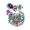
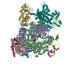
| Entry | Database: PDB / ID: 6vnw | ||||||
|---|---|---|---|---|---|---|---|
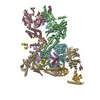
| Title | Cryo-EM structure of apo-BBSome | ||||||
 Components Components |
| ||||||
 Keywords Keywords | PROTEIN TRANSPORT | ||||||
| Function / homology |  Function and homology information Function and homology informationestablishment of anatomical structure orientation / multi-ciliated epithelial cell differentiation / receptor localization to non-motile cilium / BBSome-mediated cargo-targeting to cilium / BBSome / camera-type eye photoreceptor cell differentiation / renal tubule development / smoothened binding / establishment of planar polarity / inner ear receptor cell stereocilium organization ...establishment of anatomical structure orientation / multi-ciliated epithelial cell differentiation / receptor localization to non-motile cilium / BBSome-mediated cargo-targeting to cilium / BBSome / camera-type eye photoreceptor cell differentiation / renal tubule development / smoothened binding / establishment of planar polarity / inner ear receptor cell stereocilium organization / photoreceptor connecting cilium / patched binding / olfactory bulb development / protein localization to cilium / establishment of epithelial cell apical/basal polarity / phosphatidylinositol-3-phosphate binding / regulation of stress fiber assembly / non-motile cilium assembly / motile cilium / non-motile cilium / centrosome cycle / sensory perception / ciliary membrane / erythrocyte homeostasis / eating behavior / pericentriolar material / ciliary transition zone / B cell homeostasis / cilium assembly / axoneme / fat cell differentiation / axon guidance / protein localization to plasma membrane / multicellular organism growth / fibrillar center / Wnt signaling pathway / centriolar satellite / sensory perception of smell / intracellular protein localization / regulation of protein localization / protein transport / gene expression / RNA polymerase II-specific DNA-binding transcription factor binding / protein-macromolecule adaptor activity / neuron projection / cilium / ciliary basal body / centrosome / membrane / cytoplasm Similarity search - Function | ||||||
| Biological species |  | ||||||
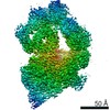
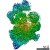
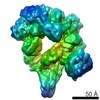
| Method | ELECTRON MICROSCOPY / single particle reconstruction / cryo EM / Resolution: 3.44 Å | ||||||
 Authors Authors | Yang, S. / Walz, T. / Nachury, M. / Chou, H. | ||||||
| Funding support |  United States, 1items United States, 1items
| ||||||
 Citation Citation | Journal: Elife / Year: 2020 Title: Near-atomic structures of the BBSome reveal the basis for BBSome activation and binding to GPCR cargoes. Authors: Shuang Yang / Kriti Bahl / Hui-Ting Chou / Jonathan Woodsmith / Ulrich Stelzl / Thomas Walz / Maxence V Nachury /  Abstract: Dynamic trafficking of G protein-coupled receptors (GPCRs) out of cilia is mediated by the BBSome. In concert with its membrane recruitment factor, the small GTPase ARL6/BBS3, the BBSome ferries ...Dynamic trafficking of G protein-coupled receptors (GPCRs) out of cilia is mediated by the BBSome. In concert with its membrane recruitment factor, the small GTPase ARL6/BBS3, the BBSome ferries GPCRs across the transition zone, a diffusion barrier at the base of cilia. Here, we present the near-atomic structures of the BBSome by itself and in complex with ARL6, and we describe the changes in BBSome conformation induced by ARL6 binding. Modeling the interactions of the BBSome with membranes and the GPCR Smoothened (SMO) reveals that SMO, and likely also other GPCR cargoes, must release their amphipathic helix 8 from the membrane to be recognized by the BBSome. | ||||||
| History |
|
- Structure visualization
| Movie |
Movie viewer |
|---|---|
| Structure viewer | Molecule: MolmilJmol/JSmol |
- Downloads & links
-Download
| PDBx/mmCIF format | 6vnw.cif.gz | 643.6 KB | Display | PDBx/mmCIF format |
|---|---|---|---|---|
| PDB format | pdb6vnw.ent.gz | 502.5 KB | Display | PDB format |
| PDBx/mmJSON format | 6vnw.json.gz | Tree view | PDBx/mmJSON format | |
| Others |  Other downloads Other downloads |
-Validation report
| Arichive directory | https://data.pdbj.org/pub/pdb/validation_reports/vn/6vnwftp://data.pdbj.org/pub/pdb/validation_reports/vn/6vnw | HTTPS FTP |
|---|
-Related structure data
| Related structure data |  21251MC  6voaC M: map data used to model this data C: citing same article ( |
|---|---|
| Similar structure data |


-Links
 PDBj
PDBj


- Assembly
| Deposited unit | 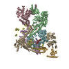
|
|---|---|
| 1 |
|
-Components
-Bardet-Biedl syndrome ... , 6 types, 6 molecules HBEGCI
| #1: Protein | Mass: 8070.502 Da / Num. of mol.: 1 / Source method: isolated from a natural source / Source: (natural) |
|---|---|
| #2: Protein | Mass: 79911.484 Da / Num. of mol.: 1 / Source method: isolated from a natural source / Source: (natural) |
| #3: Protein | Mass: 58289.133 Da / Num. of mol.: 1 / Source method: isolated from a natural source / Source: (natural) |
| #4: Protein | Mass: 38880.984 Da / Num. of mol.: 1 / Source method: isolated from a natural source / Source: (natural) |
| #5: Protein | Mass: 80471.375 Da / Num. of mol.: 1 / Source method: isolated from a natural source / Source: (natural) |
| #7: Protein | Mass: 99230.914 Da / Num. of mol.: 1 / Source method: isolated from a natural source / Source: (natural) |
-Protein , 2 types, 2 molecules FD
| #6: Protein | Mass: 56686.406 Da / Num. of mol.: 1 / Source method: isolated from a natural source / Source: (natural) |
|---|---|
| #8: Protein | Mass: 64939.141 Da / Num. of mol.: 1 / Source method: isolated from a natural source / Source: (natural) |
-Experimental details
-Experiment
| Experiment | Method: ELECTRON MICROSCOPY |
|---|---|
| EM experiment | Aggregation state: PARTICLE / 3D reconstruction method: single particle reconstruction |
- Sample preparation
| Component | Name: BBSome complex / Type: COMPLEX / Entity ID: all / Source: NATURAL | |||||||||||||||||||||||||
|---|---|---|---|---|---|---|---|---|---|---|---|---|---|---|---|---|---|---|---|---|---|---|---|---|---|---|
| Molecular weight | Value: 0.5 MDa / Experimental value: NO | |||||||||||||||||||||||||
| Source (natural) | Organism: | |||||||||||||||||||||||||
| Buffer solution | pH: 7.5 | |||||||||||||||||||||||||
| Buffer component |
| |||||||||||||||||||||||||
| Specimen | Conc.: 0.4 mg/ml / Embedding applied: NO / Shadowing applied: NO / Staining applied: NO / Vitrification applied: YES | |||||||||||||||||||||||||
| Specimen support | Grid material: COPPER / Grid mesh size: 400 divisions/in. / Grid type: Quantifoil R1.2/1.3 | |||||||||||||||||||||||||
| Vitrification | Instrument: FEI VITROBOT MARK IV / Cryogen name: ETHANE / Humidity: 100 % / Chamber temperature: 277 K / Details: Wait time 20s, blot force -2, blot time 3.5s |
- Electron microscopy imaging
| Experimental equipment |  Model: Titan Krios / Image courtesy: FEI Company |
|---|---|
| Microscopy | Model: FEI TITAN KRIOS |
| Electron gun | Electron source:  FIELD EMISSION GUN / Accelerating voltage: 300 kV / Illumination mode: FLOOD BEAM FIELD EMISSION GUN / Accelerating voltage: 300 kV / Illumination mode: FLOOD BEAM |
| Electron lens | Mode: BRIGHT FIELD |
| Image recording | Electron dose: 80 e/Å2 / Detector mode: SUPER-RESOLUTION / Film or detector model: GATAN K2 SUMMIT (4k x 4k) |
- Processing
| EM software |
| ||||||||||||
|---|---|---|---|---|---|---|---|---|---|---|---|---|---|
| CTF correction | Type: PHASE FLIPPING AND AMPLITUDE CORRECTION | ||||||||||||
| Symmetry | Point symmetry: C1 (asymmetric) | ||||||||||||
| 3D reconstruction | Resolution: 3.44 Å / Resolution method: FSC 0.143 CUT-OFF / Num. of particles: 560777 / Symmetry type: POINT |
