 Movie
Movie Controller

[English] 日本語
 Yorodumi
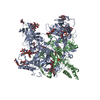
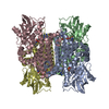
- PDB-6vkm: Crystal Structure of Stabilized GP from Makona Variant of Ebola Virus -
+ Open data

- Basic information
| Entry | Database: PDB / ID: 6vkm | |||||||||
|---|---|---|---|---|---|---|---|---|---|---|
| Title | Crystal Structure of Stabilized GP from Makona Variant of Ebola Virus | |||||||||
 Components Components | Virion spike glycoprotein | |||||||||
 Keywords Keywords | VIRAL PROTEIN / virus / glycoprotein / fusion / trimer / prefusion / EBOV | |||||||||
| Function / homology |  Function and homology information Function and homology informationsymbiont-mediated killing of host cell / host cell endoplasmic reticulum / viral budding from plasma membrane / symbiont-mediated-mediated suppression of host tetherin activity / clathrin-dependent endocytosis of virus by host cell / host cell cytoplasm / entry receptor-mediated virion attachment to host cell / symbiont-mediated suppression of host innate immune response / membrane raft / fusion of virus membrane with host endosome membrane ...symbiont-mediated killing of host cell / host cell endoplasmic reticulum / viral budding from plasma membrane / symbiont-mediated-mediated suppression of host tetherin activity / clathrin-dependent endocytosis of virus by host cell / host cell cytoplasm / entry receptor-mediated virion attachment to host cell / symbiont-mediated suppression of host innate immune response / membrane raft / fusion of virus membrane with host endosome membrane / viral envelope / symbiont entry into host cell / lipid binding / host cell plasma membrane / virion membrane / extracellular region / membrane / identical protein binding Similarity search - Function | |||||||||
| Biological species |   Zaire ebolavirus Zaire ebolavirus | |||||||||
| Method |  X-RAY DIFFRACTION / SYNCHROTRON / MOLECULAR REPLACEMENT / Resolution: 3.5 Å X-RAY DIFFRACTION / SYNCHROTRON / MOLECULAR REPLACEMENT / Resolution: 3.5 Å | |||||||||
 Authors Authors | Gilman, M.S.A. / Rutten, L. / Langedijk, J.P.M. / McLellan, J.S. | |||||||||
 Citation Citation | Journal: Cell Rep / Year: 2020 Title: Structure-Based Design of Prefusion-Stabilized Filovirus Glycoprotein Trimers. Authors: Rutten, L. / Gilman, M.S.A. / Blokland, S. / Juraszek, J. / McLellan, J.S. / Langedijk, J.P.M. | |||||||||
| History |
|
- Structure visualization
| Structure viewer | Molecule: MolmilJmol/JSmol |
|---|
- Downloads & links
-Download
| PDBx/mmCIF format | 6vkm.cif.gz | 157.6 KB | Display | PDBx/mmCIF format |
|---|---|---|---|---|
| PDB format | pdb6vkm.ent.gz | 122.6 KB | Display | PDB format |
| PDBx/mmJSON format | 6vkm.json.gz | Tree view | PDBx/mmJSON format | |
| Others |  Other downloads Other downloads |
-Validation report
| Arichive directory | https://data.pdbj.org/pub/pdb/validation_reports/vk/6vkmftp://data.pdbj.org/pub/pdb/validation_reports/vk/6vkm | HTTPS FTP |
|---|
-Related structure data
| Related structure data |  5jq3S S: Starting model for refinement |
|---|---|
| Similar structure data |


-Links
 PDBj
PDBj

- Assembly
| Deposited unit | 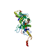
| ||||||||
|---|---|---|---|---|---|---|---|---|---|
| 1 | 
| ||||||||
| Unit cell |
|
-Components
| #1: Protein | Mass: 55446.449 Da / Num. of mol.: 1 / Mutation: T42A/T577P/K588F,deletion of residues 320-476 Source method: isolated from a genetically manipulated source Source: (gene. exp.) Zaire ebolavirus / Gene: GP / Cell line (production host): Expi293F / Production host:  Homo sapiens (human) Homo sapiens (human)References: UniProt: A0A2I4PEY8, UniProt: A0A0E3XL33, UniProt: Q05320*PLUS |
|---|---|
| #2: Polysaccharide | 2-acetamido-2-deoxy-beta-D-glucopyranose-(1-4)-2-acetamido-2-deoxy-beta-D-glucopyranose Source method: isolated from a genetically manipulated source |
| #3: Sugar | ChemComp-NAG /   Type: D-saccharide, beta linking / Mass: 221.208 Da / Num. of mol.: 1 Type: D-saccharide, beta linking / Mass: 221.208 Da / Num. of mol.: 1Source method: isolated from a genetically manipulated source Formula: C8H15NO6 |
| Has ligand of interest | N |
| Has protein modification | Y |
-Experimental details
-Experiment
| Experiment | Method: X-RAY DIFFRACTION / Number of used crystals: 1 |
|---|
- Sample preparation
| Crystal | Density Matthews: 4.36 Å3/Da / Density % sol: 71.82 % |
|---|---|
| Crystal grow | Temperature: 293 K / Method: vapor diffusion, hanging drop / pH: 5.2 Details: 9.8% PEG 6000, 0.1 M Sodium citrate pH 5.2, 3% glycerol |
-Data collection
| Diffraction | Mean temperature: 80 K / Serial crystal experiment: N | ||||||||||||||||||||||||||||||
|---|---|---|---|---|---|---|---|---|---|---|---|---|---|---|---|---|---|---|---|---|---|---|---|---|---|---|---|---|---|---|---|
| Diffraction source | Source: SYNCHROTRON / Site: APS  / Beamline: 19-ID / Wavelength: 0.97918 Å / Beamline: 19-ID / Wavelength: 0.97918 Å | ||||||||||||||||||||||||||||||
| Detector | Type: DECTRIS PILATUS3 X 6M / Detector: PIXEL / Date: Oct 26, 2017 | ||||||||||||||||||||||||||||||
| Radiation | Protocol: SINGLE WAVELENGTH / Monochromatic (M) / Laue (L): M / Scattering type: x-ray | ||||||||||||||||||||||||||||||
| Radiation wavelength | Wavelength: 0.97918 Å / Relative weight: 1 | ||||||||||||||||||||||||||||||
| Reflection | Resolution: 3.5→47.53 Å / Num. obs: 10491 / % possible obs: 84.7 % / Redundancy: 4.3 % / CC1/2: 0.913 / Rmerge(I) obs: 0.346 / Rpim(I) all: 0.177 / Rrim(I) all: 0.391 / Net I/σ(I): 3.4 / Num. measured all: 45250 / Scaling rejects: 3 | ||||||||||||||||||||||||||||||
| Reflection shell | Diffraction-ID: 1
|
- Processing
| Software |
| |||||||||||||||||||||||||||||||||||||||||||||||||||||||||||||||||||||||||||
|---|---|---|---|---|---|---|---|---|---|---|---|---|---|---|---|---|---|---|---|---|---|---|---|---|---|---|---|---|---|---|---|---|---|---|---|---|---|---|---|---|---|---|---|---|---|---|---|---|---|---|---|---|---|---|---|---|---|---|---|---|---|---|---|---|---|---|---|---|---|---|---|---|---|---|---|---|
| Refinement | Method to determine structure: MOLECULAR REPLACEMENT Starting model: 5JQ3 Resolution: 3.5→47.53 Å / SU ML: 0.53 / Cross valid method: THROUGHOUT / σ(F): 1.34 / Phase error: 30.89
| |||||||||||||||||||||||||||||||||||||||||||||||||||||||||||||||||||||||||||
| Solvent computation | Shrinkage radii: 0.9 Å / VDW probe radii: 1.11 Å | |||||||||||||||||||||||||||||||||||||||||||||||||||||||||||||||||||||||||||
| Displacement parameters | Biso max: 130.14 Å2 / Biso mean: 62.1766 Å2 / Biso min: 33.85 Å2 | |||||||||||||||||||||||||||||||||||||||||||||||||||||||||||||||||||||||||||
| Refinement step | Cycle: final / Resolution: 3.5→47.53 Å
| |||||||||||||||||||||||||||||||||||||||||||||||||||||||||||||||||||||||||||
| LS refinement shell | Refine-ID: X-RAY DIFFRACTION / Rfactor Rfree error: 0
| |||||||||||||||||||||||||||||||||||||||||||||||||||||||||||||||||||||||||||
| Refinement TLS params. | Method: refined / Refine-ID: X-RAY DIFFRACTION
| |||||||||||||||||||||||||||||||||||||||||||||||||||||||||||||||||||||||||||
| Refinement TLS group |
|
