 Movie
Movie Controller

[English] 日本語
 Yorodumi
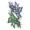
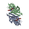
- PDB-6nae: Crystal Structure of Ebola zaire GP protein with bound ARN0074898 -
+ Open data

- Basic information
| Entry | Database: PDB / ID: 6nae | |||||||||
|---|---|---|---|---|---|---|---|---|---|---|
| Title | Crystal Structure of Ebola zaire GP protein with bound ARN0074898 | |||||||||
 Components Components |
| |||||||||
 Keywords Keywords | VIRAL PROTEIN / SSGCID / Ebola zaire / glycoprotein / Structural Genomics / Seattle Structural Genomics Center for Infectious Disease | |||||||||
| Function / homology |  Function and homology information Function and homology informationsymbiont-mediated killing of host cell / host cell endoplasmic reticulum / viral budding from plasma membrane / symbiont-mediated-mediated suppression of host tetherin activity / clathrin-dependent endocytosis of virus by host cell / host cell cytoplasm / entry receptor-mediated virion attachment to host cell / symbiont-mediated suppression of host innate immune response / membrane raft / fusion of virus membrane with host endosome membrane ...symbiont-mediated killing of host cell / host cell endoplasmic reticulum / viral budding from plasma membrane / symbiont-mediated-mediated suppression of host tetherin activity / clathrin-dependent endocytosis of virus by host cell / host cell cytoplasm / entry receptor-mediated virion attachment to host cell / symbiont-mediated suppression of host innate immune response / membrane raft / fusion of virus membrane with host endosome membrane / viral envelope / symbiont entry into host cell / lipid binding / host cell plasma membrane / virion membrane / extracellular region / identical protein binding Similarity search - Function | |||||||||
| Biological species |   Zaire ebolavirus Zaire ebolavirus | |||||||||
| Method |  X-RAY DIFFRACTION / SYNCHROTRON / MOLECULAR REPLACEMENT / molecular replacement / Resolution: 2.75 Å X-RAY DIFFRACTION / SYNCHROTRON / MOLECULAR REPLACEMENT / molecular replacement / Resolution: 2.75 Å | |||||||||
 Authors Authors | Seattle Structural Genomics Center for Infectious Disease (SSGCID) | |||||||||
 Citation Citation | Journal: Acs Med.Chem.Lett. / Year: 2020 Title: Discovery of Adamantane Carboxamides as Ebola Virus Cell Entry and Glycoprotein Inhibitors. Authors: Plewe, M.B. / Sokolova, N.V. / Gantla, V.R. / Brown, E.R. / Naik, S. / Fetsko, A. / Lorimer, D.D. / Dranow, D.M. / Smutney, H. / Bullen, J. / Sidhu, R. / Master, A. / Wang, J. / Kallel, E.A. ...Authors: Plewe, M.B. / Sokolova, N.V. / Gantla, V.R. / Brown, E.R. / Naik, S. / Fetsko, A. / Lorimer, D.D. / Dranow, D.M. / Smutney, H. / Bullen, J. / Sidhu, R. / Master, A. / Wang, J. / Kallel, E.A. / Zhang, L. / Kalveram, B. / Freiberg, A.N. / Henkel, G. / McCormack, K. | |||||||||
| History |
|
- Structure visualization
| Structure viewer | Molecule: MolmilJmol/JSmol |
|---|
- Downloads & links
-Download
| PDBx/mmCIF format | 6nae.cif.gz | 98.6 KB | Display | PDBx/mmCIF format |
|---|---|---|---|---|
| PDB format | pdb6nae.ent.gz | 69.2 KB | Display | PDB format |
| PDBx/mmJSON format | 6nae.json.gz | Tree view | PDBx/mmJSON format | |
| Others |  Other downloads Other downloads |
-Validation report
| Arichive directory | https://data.pdbj.org/pub/pdb/validation_reports/na/6naeftp://data.pdbj.org/pub/pdb/validation_reports/na/6nae | HTTPS FTP |
|---|
-Related structure data
| Related structure data |  6f5uS S: Starting model for refinement |
|---|---|
| Similar structure data | |
| Other databases |

-Links
 PDBj
PDBj

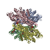
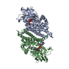
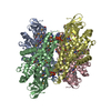
- Assembly
| Deposited unit | 
| ||||||||
|---|---|---|---|---|---|---|---|---|---|
| 1 | 
| ||||||||
| Unit cell |
|
-Components
-Protein , 2 types, 2 molecules AB
| #1: Protein | Mass: 36302.719 Da / Num. of mol.: 1 Fragment: EbzaA.19907.a.HE11,EbzaA.19907.a.HE11,EbzaA.19907.a.HE11,EbzaA.19907.a.HE11,EbzaA.19907.a.HE11,EbzaA.19907.a.HE11,EbzaA.19907.a.HE11,EbzaA.19907.a.HE11,EbzaA.19907.a.HE11 Mutation: T42A Source method: isolated from a genetically manipulated source Source: (gene. exp.) Zaire ebolavirus (strain Mayinga-76) / Strain: Mayinga-76 / Gene: GP / Plasmid: EbzaA.19907.a.HE11 / Production host:  Homo sapiens (human) / Strain (production host): HEK-293 / References: UniProt: Q05320 Homo sapiens (human) / Strain (production host): HEK-293 / References: UniProt: Q05320 |
|---|---|
| #2: Protein | Mass: 18922.320 Da / Num. of mol.: 1 / Mutation: H613A Source method: isolated from a genetically manipulated source Source: (gene. exp.) Zaire ebolavirus (strain Mayinga-76) / Strain: Mayinga-76 / Gene: GP / Plasmid: EbzaA.19907.a.HE11 / Production host: Homo sapiens (human) / Strain (production host): HEK-293 / References: UniProt: Q05320 |
-Sugars , 2 types, 5 molecules 
| #3: Polysaccharide | alpha-D-mannopyranose-(1-3)-[alpha-D-mannopyranose-(1-6)]beta-D-mannopyranose-(1-4)-2-acetamido-2- ...alpha-D-mannopyranose-(1-3)-[alpha-D-mannopyranose-(1-6)]beta-D-mannopyranose-(1-4)-2-acetamido-2-deoxy-beta-D-glucopyranose-(1-4)-2-acetamido-2-deoxy-beta-D-glucopyranose Source method: isolated from a genetically manipulated source |
|---|---|
| #4: Sugar | ChemComp-NAG /  Type: D-saccharide, beta linking / Mass: 221.208 Da / Num. of mol.: 4 / Source method: obtained synthetically / Formula: C8H15NO6 Type: D-saccharide, beta linking / Mass: 221.208 Da / Num. of mol.: 4 / Source method: obtained synthetically / Formula: C8H15NO6 |
-Non-polymers , 3 types, 94 molecules 


| #5: Chemical | ChemComp-GOL /  Mass: 92.094 Da / Num. of mol.: 5 / Source method: obtained synthetically / Formula: C3H8O3 Mass: 92.094 Da / Num. of mol.: 5 / Source method: obtained synthetically / Formula: C3H8O3#6: Chemical | ChemComp-KHG / ( |  Mass: 366.540 Da / Num. of mol.: 1 Mass: 366.540 Da / Num. of mol.: 1Source method: isolated from a genetically manipulated source Formula: C24H34N2O #7: Water | ChemComp-HOH / | Mass: 18.015 Da / Num. of mol.: 88 / Source method: isolated from a natural source / Formula: H2O |
|---|
-Details
| Has protein modification | Y |
|---|
-Experimental details
-Experiment
| Experiment | Method: X-RAY DIFFRACTION / Number of used crystals: 1 |
|---|
- Sample preparation
| Crystal | Density Matthews: 3.47 Å3/Da / Density % sol: 64.55 % |
|---|---|
| Crystal grow | Temperature: 290 K / Method: vapor diffusion, sitting drop / pH: 7.1 Details: EbzaA.19907.a.HE11.PD38326 at 6.01 mg /ml and mixed 1:1 with an opt screen based on JCSG+(b8): 11 % (w/v) PEG-8000, 0.1 M Tris base/ HCl, pH = 7.1, 200 mM MgCl2, Crystals were soaked with 1 ...Details: EbzaA.19907.a.HE11.PD38326 at 6.01 mg /ml and mixed 1:1 with an opt screen based on JCSG+(b8): 11 % (w/v) PEG-8000, 0.1 M Tris base/ HCl, pH = 7.1, 200 mM MgCl2, Crystals were soaked with 1 mM ARN0074898 for 4 hours and cryoprotected with 20% glyerol. Tray: 303411b4, puck: jvo4-3 |
-Data collection
| Diffraction | Mean temperature: 100 K / Serial crystal experiment: N | ||||||||||||||||||||||||||||||||||||||||||||||||||||||||||||||||||||||||||||||||||||||||||||||||||||||||||||||||||||||||||||||||||||||||||||||||||||||||||||||||||||||||
|---|---|---|---|---|---|---|---|---|---|---|---|---|---|---|---|---|---|---|---|---|---|---|---|---|---|---|---|---|---|---|---|---|---|---|---|---|---|---|---|---|---|---|---|---|---|---|---|---|---|---|---|---|---|---|---|---|---|---|---|---|---|---|---|---|---|---|---|---|---|---|---|---|---|---|---|---|---|---|---|---|---|---|---|---|---|---|---|---|---|---|---|---|---|---|---|---|---|---|---|---|---|---|---|---|---|---|---|---|---|---|---|---|---|---|---|---|---|---|---|---|---|---|---|---|---|---|---|---|---|---|---|---|---|---|---|---|---|---|---|---|---|---|---|---|---|---|---|---|---|---|---|---|---|---|---|---|---|---|---|---|---|---|---|---|---|---|---|---|---|
| Diffraction source | Source: SYNCHROTRON / Site: APS  / Beamline: 21-ID-F / Wavelength: 0.97872 Å / Beamline: 21-ID-F / Wavelength: 0.97872 Å | ||||||||||||||||||||||||||||||||||||||||||||||||||||||||||||||||||||||||||||||||||||||||||||||||||||||||||||||||||||||||||||||||||||||||||||||||||||||||||||||||||||||||
| Detector | Type: RAYONIX MX-300 / Detector: CCD / Date: Oct 5, 2018 / Details: Beryllium Lenses | ||||||||||||||||||||||||||||||||||||||||||||||||||||||||||||||||||||||||||||||||||||||||||||||||||||||||||||||||||||||||||||||||||||||||||||||||||||||||||||||||||||||||
| Radiation | Protocol: SINGLE WAVELENGTH / Monochromatic (M) / Laue (L): M / Scattering type: x-ray | ||||||||||||||||||||||||||||||||||||||||||||||||||||||||||||||||||||||||||||||||||||||||||||||||||||||||||||||||||||||||||||||||||||||||||||||||||||||||||||||||||||||||
| Radiation wavelength | Wavelength: 0.97872 Å / Relative weight: 1 | ||||||||||||||||||||||||||||||||||||||||||||||||||||||||||||||||||||||||||||||||||||||||||||||||||||||||||||||||||||||||||||||||||||||||||||||||||||||||||||||||||||||||
| Reflection | Resolution: 2.75→49.554 Å / Num. obs: 20154 / % possible obs: 100 % / Redundancy: 7.409 % / Biso Wilson estimate: 57.765 Å2 / CC1/2: 0.999 / Rmerge(I) obs: 0.065 / Rrim(I) all: 0.07 / Χ2: 1.038 / Net I/σ(I): 23.09 / Num. measured all: 149327 | ||||||||||||||||||||||||||||||||||||||||||||||||||||||||||||||||||||||||||||||||||||||||||||||||||||||||||||||||||||||||||||||||||||||||||||||||||||||||||||||||||||||||
| Reflection shell | Diffraction-ID: 1
|
-Phasing
| Phasing | Method: molecular replacement |
|---|
- Processing
| Software |
| ||||||||||||||||||||||||||||||||||||||||||||||||||||||||||||||||||||||||||||||||||||||||||||||||
|---|---|---|---|---|---|---|---|---|---|---|---|---|---|---|---|---|---|---|---|---|---|---|---|---|---|---|---|---|---|---|---|---|---|---|---|---|---|---|---|---|---|---|---|---|---|---|---|---|---|---|---|---|---|---|---|---|---|---|---|---|---|---|---|---|---|---|---|---|---|---|---|---|---|---|---|---|---|---|---|---|---|---|---|---|---|---|---|---|---|---|---|---|---|---|---|---|---|
| Refinement | Method to determine structure: MOLECULAR REPLACEMENT Starting model: 6f5u Resolution: 2.75→49.554 Å / SU ML: 0.33 / Cross valid method: FREE R-VALUE / σ(F): 1.36 / Phase error: 23.98
| ||||||||||||||||||||||||||||||||||||||||||||||||||||||||||||||||||||||||||||||||||||||||||||||||
| Solvent computation | Shrinkage radii: 0.9 Å / VDW probe radii: 1.11 Å | ||||||||||||||||||||||||||||||||||||||||||||||||||||||||||||||||||||||||||||||||||||||||||||||||
| Displacement parameters | Biso max: 168.95 Å2 / Biso mean: 65.2952 Å2 / Biso min: 26.28 Å2 | ||||||||||||||||||||||||||||||||||||||||||||||||||||||||||||||||||||||||||||||||||||||||||||||||
| Refinement step | Cycle: final / Resolution: 2.75→49.554 Å
| ||||||||||||||||||||||||||||||||||||||||||||||||||||||||||||||||||||||||||||||||||||||||||||||||
| LS refinement shell | Refine-ID: X-RAY DIFFRACTION / Rfactor Rfree error: 0 / Total num. of bins used: 15 / % reflection obs: 100 %
|
