 Movie
Movie Controller

[English] 日本語
 Yorodumi
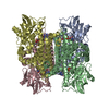
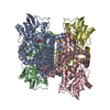
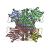
- PDB-3d9g: Nitroalkane oxidase: wild type crystallized in a trapped state fo... -
+ Open data

- Basic information
| Entry | Database: PDB / ID: 3d9g | ||||||
|---|---|---|---|---|---|---|---|
| Title | Nitroalkane oxidase: wild type crystallized in a trapped state forming a cyanoadduct with FAD | ||||||
 Components Components | Nitroalkane oxidase | ||||||
 Keywords Keywords | OXIDOREDUCTASE / FLAVOENZYME / NITROALKANE / ACYL-COA DEHYDROGENASE / LONG CELL EDGE / FAD / INHIBITOR / FLAVOPROTEIN | ||||||
| Function / homology |  Function and homology information Function and homology informationnitroalkane oxidase / nitroalkane oxidase activity / butyrate catabolic process / detoxification / oxidoreductase activity, acting on the CH-CH group of donors / fatty acid beta-oxidation using acyl-CoA dehydrogenase / FAD binding Similarity search - Function | ||||||
| Biological species |   Fusarium oxysporum (fungus) Fusarium oxysporum (fungus) | ||||||
| Method |  X-RAY DIFFRACTION / SYNCHROTRON / MOLECULAR REPLACEMENT / Resolution: 2.15 Å X-RAY DIFFRACTION / SYNCHROTRON / MOLECULAR REPLACEMENT / Resolution: 2.15 Å | ||||||
 Authors Authors | Heroux, A. / Bozinovski, D.M. / Valley, M.P. / Fitzpatrick, P.F. / Orville, A.M. | ||||||
 Citation Citation | Journal: Biochemistry / Year: 2009 Title: Crystal structures of intermediates in the nitroalkane oxidase reaction. Authors: Heroux, A. / Bozinovski, D.M. / Valley, M.P. / Fitzpatrick, P.F. / Orville, A.M. #1: Journal: Biochemistry / Year: 2006Title: Crystal structures of nitroalkane oxidase: insights into the reaction mechanism from a covalent complex of the flavoenzyme trapped during turnover. Authors: Nagpal, A. / Valley, M.P. / Fitzpatrick, P.F. / Orville, A.M. #2: Journal: Biochemistry / Year: 2007Title: Mechanistic and structural analyses of the roles of Arg409 and Asp402 in the reaction of the flavoprotein nitroalkane oxidase. Authors: Fitzpatrick, P.F. / Bozinovski, D.M. / Heroux, A. / Shaw, P.G. / Valley, M.P. / Orville, A.M. | ||||||
| History |
|
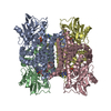
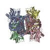
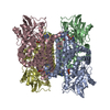
- Structure visualization
| Structure viewer | Molecule: MolmilJmol/JSmol |
|---|
- Downloads & links
-Download
| PDBx/mmCIF format | 3d9g.cif.gz | 351.8 KB | Display | PDBx/mmCIF format |
|---|---|---|---|---|
| PDB format | pdb3d9g.ent.gz | 286.4 KB | Display | PDB format |
| PDBx/mmJSON format | 3d9g.json.gz | Tree view | PDBx/mmJSON format | |
| Others |  Other downloads Other downloads |
-Validation report
| Arichive directory | https://data.pdbj.org/pub/pdb/validation_reports/d9/3d9gftp://data.pdbj.org/pub/pdb/validation_reports/d9/3d9g | HTTPS FTP |
|---|
-Related structure data
| Related structure data |  3d9dC  3d9eC  3d9fC  2c12S S: Starting model for refinement C: citing same article ( |
|---|---|
| Similar structure data |


-Links
 PDBj
PDBj- Assembly
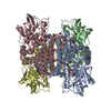
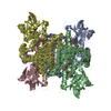
| Deposited unit | 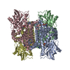
| ||||||||
|---|---|---|---|---|---|---|---|---|---|
| 1 |
| ||||||||
| Unit cell |
|
-Components
| #1: Protein | Mass: 48088.051 Da / Num. of mol.: 4 Source method: isolated from a genetically manipulated source Source: (gene. exp.) Fusarium oxysporum (fungus) / Plasmid: PETNAO4 / Production host:  #2: Chemical | ChemComp-FAD /   Mass: 785.550 Da / Num. of mol.: 4 / Source method: obtained synthetically / Formula: C27H33N9O15P2 / Comment: FAD*YM Mass: 785.550 Da / Num. of mol.: 4 / Source method: obtained synthetically / Formula: C27H33N9O15P2 / Comment: FAD*YM#3: Chemical | ChemComp-CNX /   Mass: 111.185 Da / Num. of mol.: 4 / Source method: obtained synthetically / Formula: C7H13N Mass: 111.185 Da / Num. of mol.: 4 / Source method: obtained synthetically / Formula: C7H13N#4: Chemical |   Mass: 92.094 Da / Num. of mol.: 2 / Source method: obtained synthetically / Formula: C3H8O3 Mass: 92.094 Da / Num. of mol.: 2 / Source method: obtained synthetically / Formula: C3H8O3#5: Water | ChemComp-HOH / |  Mass: 18.015 Da / Num. of mol.: 840 / Source method: isolated from a natural source / Formula: H2O Mass: 18.015 Da / Num. of mol.: 840 / Source method: isolated from a natural source / Formula: H2O |
|---|
-Experimental details
-Experiment
| Experiment | Method: X-RAY DIFFRACTION / Number of used crystals: 1 |
|---|
- Sample preparation
| Crystal | Density Matthews: 3.08 Å3/Da / Density % sol: 60.07 % |
|---|---|
| Crystal grow | Temperature: 298 K / Method: vapor diffusion, hanging drop / pH: 7.5 Details: 20-30mM PEG 3350, 20-35% glycerol, 0.1M NaCacodylate, pH 7.50, vapor diffusion, hanging drop, temperature 298K |
-Data collection
| Diffraction | Mean temperature: 100 K |
|---|---|
| Diffraction source | Source: SYNCHROTRON / Site: NSLS  / Beamline: X29A / Wavelength: 1.0809 Å / Beamline: X29A / Wavelength: 1.0809 Å |
| Detector | Type: ADSC QUANTUM 315 / Detector: CCD / Date: Apr 13, 2007 / Details: mirrors |
| Radiation | Monochromator: SAGITALLY FOCUSED Si(111) / Protocol: SINGLE WAVELENGTH / Monochromatic (M) / Laue (L): M / Scattering type: x-ray |
| Radiation wavelength | Wavelength: 1.0809 Å / Relative weight: 1 |
| Reflection | Resolution: 2.15→50 Å / Num. obs: 122023 / % possible obs: 94.2 % / Observed criterion σ(F): -3 / Observed criterion σ(I): 0 / Redundancy: 6.6 % / Rmerge(I) obs: 0.104 / Rsym value: 0.104 / Net I/σ(I): 27.4 |
| Reflection shell | Resolution: 2.15→2.23 Å / Redundancy: 1.8 % / Rmerge(I) obs: 0.304 / Mean I/σ(I) obs: 1.9 / Num. unique all: 8294 / Rsym value: 0.304 / % possible all: 64.9 |
- Processing
| Software |
| ||||||||||||||||||||||||||||||||||||||||||||||||||||||||||||||||||||||||||||||||||||||||||
|---|---|---|---|---|---|---|---|---|---|---|---|---|---|---|---|---|---|---|---|---|---|---|---|---|---|---|---|---|---|---|---|---|---|---|---|---|---|---|---|---|---|---|---|---|---|---|---|---|---|---|---|---|---|---|---|---|---|---|---|---|---|---|---|---|---|---|---|---|---|---|---|---|---|---|---|---|---|---|---|---|---|---|---|---|---|---|---|---|---|---|---|
| Refinement | Method to determine structure: MOLECULAR REPLACEMENT Starting model: 2C12 Resolution: 2.15→50 Å / Cor.coef. Fo:Fc: 0.954 / Cor.coef. Fo:Fc free: 0.941 / SU B: 4.821 / SU ML: 0.125 / Cross valid method: THROUGHOUT / σ(F): 0 / ESU R: 0.216 / ESU R Free: 0.183 / Stereochemistry target values: MAXIMUM LIKELIHOOD
| ||||||||||||||||||||||||||||||||||||||||||||||||||||||||||||||||||||||||||||||||||||||||||
| Solvent computation | Ion probe radii: 0.8 Å / Shrinkage radii: 0.8 Å / VDW probe radii: 1.4 Å / Solvent model: BABINET MODEL WITH MASK | ||||||||||||||||||||||||||||||||||||||||||||||||||||||||||||||||||||||||||||||||||||||||||
| Displacement parameters | Biso mean: 38.046 Å2
| ||||||||||||||||||||||||||||||||||||||||||||||||||||||||||||||||||||||||||||||||||||||||||
| Refinement step | Cycle: LAST / Resolution: 2.15→50 Å
| ||||||||||||||||||||||||||||||||||||||||||||||||||||||||||||||||||||||||||||||||||||||||||
| Refine LS restraints |
| ||||||||||||||||||||||||||||||||||||||||||||||||||||||||||||||||||||||||||||||||||||||||||
| LS refinement shell | Resolution: 2.154→2.21 Å / Total num. of bins used: 20
|
