- PDB-2rij: Crystal structure of a putative 2,3,4,5-tetrahydropyridine-2-carb... -
+
Open data
ID or keywords:
Loading...
-
Basic information
Entry
Database: PDB / ID: 2rij
Title
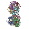
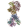
Crystal structure of a putative 2,3,4,5-tetrahydropyridine-2-carboxylate n-succinyltransferase (cj1605c, dapd) from campylobacter jejuni at 1.90 A resolution
SEQUENCE THE CONSTRUCT WAS EXPRESSED WITH A PURIFICATION TAG MGSDKIHHHHHHENLYFQG. THE TAG WAS ... SEQUENCE THE CONSTRUCT WAS EXPRESSED WITH A PURIFICATION TAG MGSDKIHHHHHHENLYFQG. THE TAG WAS REMOVED WITH TEV PROTEASE LEAVING ONLY A GLYCINE (0) FOLLOWED BY THE TARGET SEQUENCE.
SIZE EXCLUSION CHROMATOGRAPHY WITH STATIC LIGHT SCATTERING SUPPORTS THE ASSIGNMENT OF BOTH HEXAMER AND TRIMER AS SIGNIFICANT OLIGOMERIZATION STATES IN SOLUTION.
Resolution: 1.9→29.54 Å / Num. obs: 59121 / % possible obs: 100 % / Redundancy: 7.5 % / Biso Wilson estimate: 26.2 Å2 / Rmerge(I) obs: 0.124 / Rsym value: 0.124 / Net I/σ(I): 4.1
Reflection shell
Rmerge(I) obs: 0.011 / Diffraction-ID: 1
Resolution (Å)
Redundancy (%)
Mean I/σ(I) obs
Num. measured all
Num. unique all
Rsym value
% possible all
1.9-1.95
7.5
0.7
32463
4335
1.068
100
1.95-2
7.5
0.9
31624
4209
0.838
100
2-2.06
7.5
1.2
30914
4112
0.643
100
2.06-2.12
7.5
1.5
30060
3997
0.508
100
2.12-2.19
7.5
2
29252
3885
0.379
100
2.19-2.27
7.5
2.3
28140
3734
0.324
100
2.27-2.36
7.5
2.7
27324
3627
0.275
100
2.36-2.45
7.5
3
26519
3518
0.246
100
2.45-2.56
7.5
3.5
25032
3320
0.208
100
2.56-2.69
7.6
4.2
24437
3233
0.175
100
2.69-2.83
7.5
5.2
22857
3028
0.14
100
2.83-3
7.5
6.1
22005
2916
0.114
100
3-3.21
7.5
6.3
20368
2715
0.105
100
3.21-3.47
7.4
6.3
18830
2537
0.095
100
3.47-3.8
7.4
7.4
17253
2346
0.081
100
3.8-4.25
7.4
8.4
15819
2131
0.07
100
4.25-4.91
7.4
7.9
13975
1884
0.064
100
4.91-6.01
7.4
7.6
11856
1608
0.071
100
6.01-8.5
7.2
8.5
9213
1272
0.068
100
8.5-29.54
6.8
6.2
4835
714
0.07
97.5
-
Phasing
Phasing
Method: MAD
-
Processing
Software
Name
Version
Classification
NB
REFMAC
5.2.0019
refinement
PHENIX
refinement
SHELX
phasing
MolProbity
3beta29
modelbuilding
SCALA
datascaling
PDB_EXTRACT
3
dataextraction
MAR345
CCD
datacollection
MOSFLM
datareduction
SHELXD
phasing
SHARP
phasing
Refinement
Method to determine structure: MAD / Resolution: 1.9→29.54 Å / Cor.coef. Fo:Fc: 0.972 / Cor.coef. Fo:Fc free: 0.959 / SU B: 4.196 / SU ML: 0.062 / TLS residual ADP flag: LIKELY RESIDUAL / Cross valid method: THROUGHOUT / σ(F): 0 / ESU R: 0.086 / ESU R Free: 0.089 Stereochemistry target values: MAXIMUM LIKELIHOOD WITH PHASES Details: 1. HYDROGENS HAVE BEEN ADDED IN THE RIDING POSITIONS. 2. A MET-INHIBITION PROTOCOL WAS USED FOR SELENOMETHIONINE INCORPORATION DURING PROTEIN EXPRESSION. THE OCCUPANCY OF THE SE ATOMS IN THE ...Details: 1. HYDROGENS HAVE BEEN ADDED IN THE RIDING POSITIONS. 2. A MET-INHIBITION PROTOCOL WAS USED FOR SELENOMETHIONINE INCORPORATION DURING PROTEIN EXPRESSION. THE OCCUPANCY OF THE SE ATOMS IN THE MSE RESIDUES WAS REDUCED TO 0.75 FOR THE REDUCED SCATTERING POWER DUE TO PARTIAL S-MET INCORPORATION. 3. ATOM RECORD CONTAINS RESIDUAL B FACTORS ONLY. 4. CITRATE, CL AND GLYCEROL ARE MODELED BASED ON CRYSTALLIZATION AND CRYO CONDITIONS. 5. THERE IS UNMODELED DENSITY NEAR ARG 218.
Rfactor
Num. reflection
% reflection
Selection details
Rfree
0.185
2991
5.1 %
RANDOM
Rwork
0.156
-
-
-
obs
0.157
59119
100 %
-
Solvent computation
Ion probe radii: 0.8 Å / Shrinkage radii: 0.8 Å / VDW probe radii: 1.2 Å / Solvent model: MASK
Displacement parameters
Biso mean: 21.932 Å2
Baniso -1
Baniso -2
Baniso -3
1-
1.28 Å2
0.64 Å2
0 Å2
2-
-
1.28 Å2
0 Å2
3-
-
-
-1.91 Å2
Refinement step
Cycle: LAST / Resolution: 1.9→29.54 Å
Protein
Nucleic acid
Ligand
Solvent
Total
Num. atoms
2904
0
21
448
3373
Refine LS restraints
Refine-ID
Type
Dev ideal
Dev ideal target
Number
X-RAY DIFFRACTION
r_bond_refined_d
0.017
0.022
3070
X-RAY DIFFRACTION
r_bond_other_d
0.001
0.02
2095
X-RAY DIFFRACTION
r_angle_refined_deg
1.55
1.989
4162
X-RAY DIFFRACTION
r_angle_other_deg
0.953
3
5173
X-RAY DIFFRACTION
r_dihedral_angle_1_deg
5.982
5
413
X-RAY DIFFRACTION
r_dihedral_angle_2_deg
37.637
25.354
127
X-RAY DIFFRACTION
r_dihedral_angle_3_deg
12.095
15
564
X-RAY DIFFRACTION
r_dihedral_angle_4_deg
18.951
15
11
X-RAY DIFFRACTION
r_chiral_restr
0.096
0.2
476
X-RAY DIFFRACTION
r_gen_planes_refined
0.007
0.02
3441
X-RAY DIFFRACTION
r_gen_planes_other
0.001
0.02
597
X-RAY DIFFRACTION
r_nbd_refined
0.196
0.2
612
X-RAY DIFFRACTION
r_nbd_other
0.187
0.2
2185
X-RAY DIFFRACTION
r_nbtor_refined
0.176
0.2
1489
X-RAY DIFFRACTION
r_nbtor_other
0.084
0.2
1702
X-RAY DIFFRACTION
r_xyhbond_nbd_refined
0.155
0.2
318
X-RAY DIFFRACTION
r_symmetry_vdw_refined
0.232
0.2
19
X-RAY DIFFRACTION
r_symmetry_vdw_other
0.278
0.2
80
X-RAY DIFFRACTION
r_symmetry_hbond_refined
0.222
0.2
41
X-RAY DIFFRACTION
r_mcbond_it
2.274
3
2093
X-RAY DIFFRACTION
r_mcbond_other
0.567
3
800
X-RAY DIFFRACTION
r_mcangle_it
3.005
5
3108
X-RAY DIFFRACTION
r_scbond_it
5.439
8
1217
X-RAY DIFFRACTION
r_scangle_it
7.341
11
1037
LS refinement shell
Resolution: 1.9→1.949 Å / Total num. of bins used: 20
Rfactor
Num. reflection
% reflection
Rfree
0.322
211
-
Rwork
0.259
4105
-
all
-
4316
-
obs
-
-
100 %
Refinement TLS params.
Method: refined / Origin x: -9.842 Å / Origin y: 59.41 Å / Origin z: -28.889 Å
11
12
13
21
22
23
31
32
33
T
-0.0112 Å2
-0.0202 Å2
0.0136 Å2
-
-0.0492 Å2
0.0056 Å2
-
-
-0.0029 Å2
L
0.0673 °2
-0.0017 °2
0.0071 °2
-
0.4448 °2
-0.2014 °2
-
-
0.4055 °2
S
-0.0298 Å °
-0.0127 Å °
-0.028 Å °
0.0249 Å °
0.0207 Å °
0.0628 Å °
0.0982 Å °
-0.0427 Å °
0.0091 Å °
+
About Yorodumi
-
News
-
Feb 9, 2022. New format data for meta-information of EMDB entries
New format data for meta-information of EMDB entries
Version 3 of the EMDB header file is now the official format.
The previous official version 1.9 will be removed from the archive.
In the structure databanks used in Yorodumi, some data are registered as the other names, "COVID-19 virus" and "2019-nCoV". Here are the details of the virus and the list of structure data.
Jan 31, 2019. EMDB accession codes are about to change! (news from PDBe EMDB page)
EMDB accession codes are about to change! (news from PDBe EMDB page)
The allocation of 4 digits for EMDB accession codes will soon come to an end. Whilst these codes will remain in use, new EMDB accession codes will include an additional digit and will expand incrementally as the available range of codes is exhausted. The current 4-digit format prefixed with “EMD-” (i.e. EMD-XXXX) will advance to a 5-digit format (i.e. EMD-XXXXX), and so on. It is currently estimated that the 4-digit codes will be depleted around Spring 2019, at which point the 5-digit format will come into force.
The EM Navigator/Yorodumi systems omit the EMD- prefix.
Related info.:Q: What is EMD? / ID/Accession-code notation in Yorodumi/EM Navigator
Yorodumi is a browser for structure data from EMDB, PDB, SASBDB, etc.
This page is also the successor to EM Navigator detail page, and also detail information page/front-end page for Omokage search.
The word "yorodu" (or yorozu) is an old Japanese word meaning "ten thousand". "mi" (miru) is to see.
Related info.:EMDB / PDB / SASBDB / Comparison of 3 databanks / Yorodumi Search / Aug 31, 2016. New EM Navigator & Yorodumi / Yorodumi Papers / Jmol/JSmol / Function and homology information / Changes in new EM Navigator and Yorodumi
 Movie
Movie Controller

 Yorodumi
 Open data
 Basic information
 Components
 Keywords
 Function and homology information
 Campylobacter jejuni (Campylobacter)
 X-RAY DIFFRACTION /
  Authors
 Citation
 Structure visualization
 Downloads & links
 Other downloads


 PDBj
PDBj


 Assembly
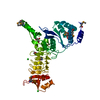


 Mass: 35.453 Da / Num. of mol.: 2 / Source method: obtained synthetically / Formula: Cl
 Mass: 192.124 Da / Num. of mol.: 1 / Source method: obtained synthetically / Formula: C6H8O7
 Mass: 92.094 Da / Num. of mol.: 1 / Source method: obtained synthetically / Formula: C3H8O3
 Mass: 18.015 Da / Num. of mol.: 448 / Source method: isolated from a natural source / Formula: H2O
 Sample preparation
 / Beamline: 23-ID-D / Wavelength: 0.97957, 0.95373, 0.97942
 Processing