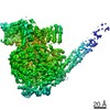
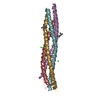
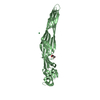
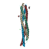
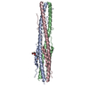
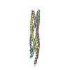
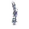
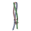
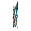
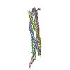
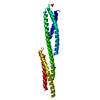
- PDB-6vag: Crystal structure of the oligomerization domain of phosphoprotein... -
+
Open data
ID or keywords:
Loading...
-
Basic information
Entry
Database: PDB / ID: 6vag
Title
Crystal structure of the oligomerization domain of phosphoprotein from parainfluenza virus 5
Components
Phosphoprotein
Keywords
VIRAL PROTEIN / Paramyxovirus / Phosphoprotein / Oligomerization domain
Function / homology
Single alpha-helices involved in coiled-coils or other helix-helix interfaces - #300 / Phosphoprotein P soyouz module / N-terminal region of Paramyxovirinae phosphoprotein (P) / P/V phosphoprotein, paramyxoviral / Paramyxovirus P/V phosphoprotein C-terminal / Single alpha-helices involved in coiled-coils or other helix-helix interfaces / Up-down Bundle / Mainly Alpha / Phosphoprotein
Journal: Proc Natl Acad Sci U S A / Year: 2020 Title: Structure of a paramyxovirus polymerase complex reveals a unique methyltransferase-CTD conformation. Authors: Ryan Abdella / Megha Aggarwal / Takashi Okura / Robert A Lamb / Yuan He / Abstract: Paramyxoviruses are enveloped, nonsegmented, negative-strand RNA viruses that cause a wide spectrum of human and animal diseases. The viral genome, packaged by the nucleoprotein (N), serves as a ...Paramyxoviruses are enveloped, nonsegmented, negative-strand RNA viruses that cause a wide spectrum of human and animal diseases. The viral genome, packaged by the nucleoprotein (N), serves as a template for the polymerase complex, composed of the large protein (L) and the homo-tetrameric phosphoprotein (P). The ∼250-kDa L possesses all enzymatic activities necessary for its function but requires P in vivo. Structural information is available for individual P domains from different paramyxoviruses, but how P interacts with L and how that affects the activity of L is largely unknown due to the lack of high-resolution structures of this complex in this viral family. In this study we determined the structure of the L-P complex from parainfluenza virus 5 (PIV5) at 4.3-Å resolution using cryoelectron microscopy, as well as the oligomerization domain (OD) of P at 1.4-Å resolution using X-ray crystallography. P-OD associates with the RNA-dependent RNA polymerase domain of L and protrudes away from it, while the X domain of one chain of P is bound near the L nucleotide entry site. The methyltransferase (MTase) domain and the C-terminal domain (CTD) of L adopt a unique conformation, positioning the MTase active site immediately above the poly-ribonucleotidyltransferase domain and near the likely exit site for the product RNA 5' end. Our study reveals a potential mechanism that mononegavirus polymerases may employ to switch between transcription and genome replication. This knowledge will assist in the design and development of antivirals against paramyxoviruses.
In the structure databanks used in Yorodumi, some data are registered as the other names, "COVID-19 virus" and "2019-nCoV". Here are the details of the virus and the list of structure data.
Jan 31, 2019. EMDB accession codes are about to change! (news from PDBe EMDB page)
EMDB accession codes are about to change! (news from PDBe EMDB page)
The allocation of 4 digits for EMDB accession codes will soon come to an end. Whilst these codes will remain in use, new EMDB accession codes will include an additional digit and will expand incrementally as the available range of codes is exhausted. The current 4-digit format prefixed with “EMD-” (i.e. EMD-XXXX) will advance to a 5-digit format (i.e. EMD-XXXXX), and so on. It is currently estimated that the 4-digit codes will be depleted around Spring 2019, at which point the 5-digit format will come into force.
The EM Navigator/Yorodumi systems omit the EMD- prefix.
Related info.:Q: What is EMD? / ID/Accession-code notation in Yorodumi/EM Navigator
Yorodumi is a browser for structure data from EMDB, PDB, SASBDB, etc.
This page is also the successor to EM Navigator detail page, and also detail information page/front-end page for Omokage search.
The word "yorodu" (or yorozu) is an old Japanese word meaning "ten thousand". "mi" (miru) is to see.
Related info.:EMDB / PDB / SASBDB / Comparison of 3 databanks / Yorodumi Search / Aug 31, 2016. New EM Navigator & Yorodumi / Yorodumi Papers / Jmol/JSmol / Function and homology information / Changes in new EM Navigator and Yorodumi
 Movie
Movie Controller

 Yorodumi
 Open data
 Basic information
 Components
 Keywords
 Function and homology information
 Parainfluenza virus 5
 X-RAY DIFFRACTION /
  Authors
 United States, 1items
  Citation
 Structure visualization
 Downloads & links
 Other downloads
 PDBj
PDBj
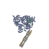
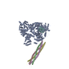
 Assembly


 Mass: 92.094 Da / Num. of mol.: 3 / Source method: obtained synthetically / Formula: C3H8O3
 Mass: 18.015 Da / Num. of mol.: 102 / Source method: isolated from a natural source / Formula: H2O
 Sample preparation
 Processing