 Movie
Movie Controller

[English] 日本語
 Yorodumi
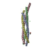
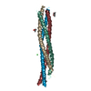
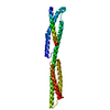
- PDB-2ztb: Crystal structure of the parasporin-2 Bacillus thuringiensis toxi... -
+ Open data

- Basic information
| Entry | Database: PDB / ID: 2ztb | ||||||
|---|---|---|---|---|---|---|---|
| Title | Crystal structure of the parasporin-2 Bacillus thuringiensis toxin that recognizes cancer cells | ||||||
 Components Components | Crystal protein | ||||||
 Keywords Keywords | TOXIN / beta-hairpin | ||||||
| Function / homology |  Function and homology information Function and homology informationImmunoglobulin-like - #3040 / Immunoglobulin-like - #4280 / Structural Genomics Hypothetical 15.5 Kd Protein In mrcA-pckA Intergenic Region; Chain A - #50 / Structural Genomics Hypothetical 15.5 Kd Protein In mrcA-pckA Intergenic Region; Chain A / Roll / Immunoglobulin-like / Sandwich / Mainly Beta / Alpha Beta Similarity search - Domain/homology | ||||||
| Biological species |  | ||||||
| Method |  X-RAY DIFFRACTION / SYNCHROTRON / MAD / Resolution: 2.38 Å X-RAY DIFFRACTION / SYNCHROTRON / MAD / Resolution: 2.38 Å | ||||||
 Authors Authors | Akiba, T. | ||||||
 Citation Citation | Journal: J.Mol.Biol. / Year: 2009 Title: Crystal structure of the parasporin-2 Bacillus thuringiensis toxin that recognizes cancer cells Authors: Akiba, T. / Abe, Y. / Kitada, S. / Kusaka, Y. / Ito, A. / Ichimatsu, T. / Katayama, H. / Akao, T. / Higuchi, K. / Mizuki, E. / Ohba, M. / Kanai, R. / Harata, K. #1: Journal: Acta Crystallogr.,Sect.D / Year: 2004 Title: Crystallization of parasporin-2, a Bacillus thuringiensis crystal protein with selective cytocidal activity against human cells Authors: Akiba, T. / Abe, Y. / Kitada, S. / Kusaka, Y. / Ito, A. / Ichimatsu, T. / Katayama, H. / Akao, T. / Higuchi, K. / Mizuki, E. / Ohba, M. / Kanai, R. / Harata, K. #2: Journal: J.Biol.Chem. / Year: 2004 Title: A Bacillus thuringiensis crystal protein with selective cytocidal action to human cells Authors: Ito, A. / Sasaguri, Y. / Kitada, S. / Kusaka, Y. / Kuwano, K. / Masutomi, K. / Mizuki, E. / Akao, T. / Ohba, M. | ||||||
| History |
|
- Structure visualization
| Structure viewer | Molecule: MolmilJmol/JSmol |
|---|
- Downloads & links
-Download
| PDBx/mmCIF format | 2ztb.cif.gz | 113.7 KB | Display | PDBx/mmCIF format |
|---|---|---|---|---|
| PDB format | pdb2ztb.ent.gz | 88.3 KB | Display | PDB format |
| PDBx/mmJSON format | 2ztb.json.gz | Tree view | PDBx/mmJSON format | |
| Others |  Other downloads Other downloads |
-Validation report
| Arichive directory | https://data.pdbj.org/pub/pdb/validation_reports/zt/2ztbftp://data.pdbj.org/pub/pdb/validation_reports/zt/2ztb | HTTPS FTP |
|---|
-Related structure data
| Similar structure data |
|---|


-Links
 PDBj
PDBj
- Assembly
| Deposited unit | 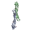
| ||||||||
|---|---|---|---|---|---|---|---|---|---|
| 1 | 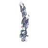
| ||||||||
| 2 | 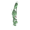
| ||||||||
| Unit cell |
|
-Components
-Protein , 1 types, 2 molecules AB
| #1: Protein | Mass: 27537.568 Da / Num. of mol.: 2 Fragment: the proteolytically activated form, UNP residues 52-302 Source method: isolated from a genetically manipulated source Source: (gene. exp.) Strain: A1547 / Plasmid: pET-23a / Production host: |
|---|
-Non-polymers , 5 types, 300 molecules 


| #2: Chemical |  Mass: 174.967 Da / Num. of mol.: 3 / Source method: obtained synthetically / Formula: Lu Mass: 174.967 Da / Num. of mol.: 3 / Source method: obtained synthetically / Formula: Lu#3: Chemical |  Mass: 92.094 Da / Num. of mol.: 2 / Source method: obtained synthetically / Formula: C3H8O3 Mass: 92.094 Da / Num. of mol.: 2 / Source method: obtained synthetically / Formula: C3H8O3#4: Chemical | ChemComp-EDO /  Mass: 62.068 Da / Num. of mol.: 4 / Source method: obtained synthetically / Formula: C2H6O2 Mass: 62.068 Da / Num. of mol.: 4 / Source method: obtained synthetically / Formula: C2H6O2#5: Chemical | ChemComp-CL / |  Mass: 35.453 Da / Num. of mol.: 1 / Source method: obtained synthetically / Formula: Cl Mass: 35.453 Da / Num. of mol.: 1 / Source method: obtained synthetically / Formula: Cl#6: Water | ChemComp-HOH / | Mass: 18.015 Da / Num. of mol.: 290 / Source method: isolated from a natural source / Formula: H2O |
|---|
-Experimental details
-Experiment
| Experiment | Method: X-RAY DIFFRACTION / Number of used crystals: 1 |
|---|
- Sample preparation
| Crystal | Density Matthews: 5.85 Å3/Da / Density % sol: 78.98 % |
|---|---|
| Crystal grow | Temperature: 298 K / Method: vapor diffusion, hanging drop / pH: 7 Details: 16% (v/v) ethylene glycol, 8% (w/v) PEG 3350, 1mM TCEP, 50mM HEPES-NaOH, pH 7.0, VAPOR DIFFUSION, HANGING DROP, temperature 298K |
-Data collection
| Diffraction | Mean temperature: 95 K | ||||||||||||
|---|---|---|---|---|---|---|---|---|---|---|---|---|---|
| Diffraction source | Source: SYNCHROTRON / Site: Photon Factory  / Beamline: BL-5A / Wavelength: 1.3403, 1.3408, 1.3460 / Beamline: BL-5A / Wavelength: 1.3403, 1.3408, 1.3460 | ||||||||||||
| Detector | Type: ADSC QUANTUM 315 / Detector: CCD / Date: Jun 11, 2004 | ||||||||||||
| Radiation | Monochromator: Si(111) double crystal monochromator / Protocol: MAD / Monochromatic (M) / Laue (L): M / Scattering type: x-ray | ||||||||||||
| Radiation wavelength |
| ||||||||||||
| Reflection | Resolution: 2.38→29.75 Å / Num. obs: 50770 / % possible obs: 99.9 % / Redundancy: 11 % / Biso Wilson estimate: 62.5 Å2 / Rmerge(I) obs: 0.067 / Net I/σ(I): 26.2 | ||||||||||||
| Reflection shell | Resolution: 2.38→2.51 Å / Redundancy: 10.6 % / Rmerge(I) obs: 0.451 / Mean I/σ(I) obs: 4.2 / Num. unique all: 7356 / % possible all: 99.8 |
- Processing
| Software |
| ||||||||||||||||||||||||||||||||||||||||||||||||||||||||||||||||||||||||||||||||||||||||||||||||||||||||||||||||||||||||||||||||||||||||||||||||||||||||||||||||||||||||||
|---|---|---|---|---|---|---|---|---|---|---|---|---|---|---|---|---|---|---|---|---|---|---|---|---|---|---|---|---|---|---|---|---|---|---|---|---|---|---|---|---|---|---|---|---|---|---|---|---|---|---|---|---|---|---|---|---|---|---|---|---|---|---|---|---|---|---|---|---|---|---|---|---|---|---|---|---|---|---|---|---|---|---|---|---|---|---|---|---|---|---|---|---|---|---|---|---|---|---|---|---|---|---|---|---|---|---|---|---|---|---|---|---|---|---|---|---|---|---|---|---|---|---|---|---|---|---|---|---|---|---|---|---|---|---|---|---|---|---|---|---|---|---|---|---|---|---|---|---|---|---|---|---|---|---|---|---|---|---|---|---|---|---|---|---|---|---|---|---|---|---|---|
| Refinement | Method to determine structure: MAD / Resolution: 2.38→29.74 Å / Cor.coef. Fo:Fc: 0.954 / Cor.coef. Fo:Fc free: 0.94 / SU B: 8.442 / SU ML: 0.112 / TLS residual ADP flag: LIKELY RESIDUAL / Cross valid method: THROUGHOUT / ESU R: 0.177 / ESU R Free: 0.165 / Stereochemistry target values: Engh & Huber / Details: HYDROGENS HAVE BEEN ADDED IN THE RIDING POSITIONS
| ||||||||||||||||||||||||||||||||||||||||||||||||||||||||||||||||||||||||||||||||||||||||||||||||||||||||||||||||||||||||||||||||||||||||||||||||||||||||||||||||||||||||||
| Solvent computation | Ion probe radii: 0.8 Å / Shrinkage radii: 0.8 Å / VDW probe radii: 1.4 Å / Solvent model: MASK | ||||||||||||||||||||||||||||||||||||||||||||||||||||||||||||||||||||||||||||||||||||||||||||||||||||||||||||||||||||||||||||||||||||||||||||||||||||||||||||||||||||||||||
| Displacement parameters | Biso mean: 58.421 Å2
| ||||||||||||||||||||||||||||||||||||||||||||||||||||||||||||||||||||||||||||||||||||||||||||||||||||||||||||||||||||||||||||||||||||||||||||||||||||||||||||||||||||||||||
| Refinement step | Cycle: LAST / Resolution: 2.38→29.74 Å
| ||||||||||||||||||||||||||||||||||||||||||||||||||||||||||||||||||||||||||||||||||||||||||||||||||||||||||||||||||||||||||||||||||||||||||||||||||||||||||||||||||||||||||
| Refine LS restraints |
| ||||||||||||||||||||||||||||||||||||||||||||||||||||||||||||||||||||||||||||||||||||||||||||||||||||||||||||||||||||||||||||||||||||||||||||||||||||||||||||||||||||||||||
| LS refinement shell | Resolution: 2.38→2.441 Å / Total num. of bins used: 20
| ||||||||||||||||||||||||||||||||||||||||||||||||||||||||||||||||||||||||||||||||||||||||||||||||||||||||||||||||||||||||||||||||||||||||||||||||||||||||||||||||||||||||||
| Refinement TLS params. | Method: refined / Refine-ID: X-RAY DIFFRACTION
| ||||||||||||||||||||||||||||||||||||||||||||||||||||||||||||||||||||||||||||||||||||||||||||||||||||||||||||||||||||||||||||||||||||||||||||||||||||||||||||||||||||||||||
| Refinement TLS group |
|
