 Movie
Movie Controller


+ Open data

- Basic information
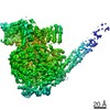
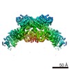
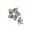
| Entry | Database: PDB / ID: 6v85 | ||||||||||||||||||
|---|---|---|---|---|---|---|---|---|---|---|---|---|---|---|---|---|---|---|---|
| Title | Parainfluenza virus 5 L-P complex | ||||||||||||||||||
 Components Components |
| ||||||||||||||||||
 Keywords Keywords | VIRAL PROTEIN / POLYMERASE / METHYLTRANSFERASE / POLY-RIBONUCLEOTIDYLTRANSFERASE | ||||||||||||||||||
| Function / homology |  Function and homology information Function and homology informationGDP polyribonucleotidyltransferase / Hydrolases; Acting on acid anhydrides; In phosphorus-containing anhydrides / virion component / host cell cytoplasm / mRNA 5'-cap (guanine-N7-)-methyltransferase activity / RNA-directed RNA polymerase / RNA-directed RNA polymerase activity / GTPase activity / ATP binding Similarity search - Function | ||||||||||||||||||
| Biological species |  Parainfluenza virus 5 Parainfluenza virus 5 | ||||||||||||||||||
| Method | ELECTRON MICROSCOPY / single particle reconstruction / cryo EM / Resolution: 4.38 Å | ||||||||||||||||||
 Authors Authors | Abdella, R. / He, Y. | ||||||||||||||||||
| Funding support |  United States, 5items United States, 5items
| ||||||||||||||||||
 Citation Citation | Journal: Proc Natl Acad Sci U S A / Year: 2020 Title: Structure of a paramyxovirus polymerase complex reveals a unique methyltransferase-CTD conformation. Authors: Ryan Abdella / Megha Aggarwal / Takashi Okura / Robert A Lamb / Yuan He / Abstract: Paramyxoviruses are enveloped, nonsegmented, negative-strand RNA viruses that cause a wide spectrum of human and animal diseases. The viral genome, packaged by the nucleoprotein (N), serves as a ...Paramyxoviruses are enveloped, nonsegmented, negative-strand RNA viruses that cause a wide spectrum of human and animal diseases. The viral genome, packaged by the nucleoprotein (N), serves as a template for the polymerase complex, composed of the large protein (L) and the homo-tetrameric phosphoprotein (P). The ∼250-kDa L possesses all enzymatic activities necessary for its function but requires P in vivo. Structural information is available for individual P domains from different paramyxoviruses, but how P interacts with L and how that affects the activity of L is largely unknown due to the lack of high-resolution structures of this complex in this viral family. In this study we determined the structure of the L-P complex from parainfluenza virus 5 (PIV5) at 4.3-Å resolution using cryoelectron microscopy, as well as the oligomerization domain (OD) of P at 1.4-Å resolution using X-ray crystallography. P-OD associates with the RNA-dependent RNA polymerase domain of L and protrudes away from it, while the X domain of one chain of P is bound near the L nucleotide entry site. The methyltransferase (MTase) domain and the C-terminal domain (CTD) of L adopt a unique conformation, positioning the MTase active site immediately above the poly-ribonucleotidyltransferase domain and near the likely exit site for the product RNA 5' end. Our study reveals a potential mechanism that mononegavirus polymerases may employ to switch between transcription and genome replication. This knowledge will assist in the design and development of antivirals against paramyxoviruses. | ||||||||||||||||||
| History |
|
- Structure visualization
| Movie |
Movie viewer |
|---|---|
| Structure viewer | Molecule: MolmilJmol/JSmol |
- Downloads & links
-Download
| PDBx/mmCIF format | 6v85.cif.gz | 436.4 KB | Display | PDBx/mmCIF format |
|---|---|---|---|---|
| PDB format | pdb6v85.ent.gz | 336.8 KB | Display | PDB format |
| PDBx/mmJSON format | 6v85.json.gz | Tree view | PDBx/mmJSON format | |
| Others |  Other downloads Other downloads |
-Validation report
| Arichive directory | https://data.pdbj.org/pub/pdb/validation_reports/v8/6v85ftp://data.pdbj.org/pub/pdb/validation_reports/v8/6v85 | HTTPS FTP |
|---|
-Related structure data
| Related structure data |  21095MC  6v86C  6vagC M: map data used to model this data C: citing same article ( |
|---|---|
| Similar structure data |

-Links
 PDBj
PDBj
- Assembly
| Deposited unit | 
|
|---|---|
| 1 |
|
-Components
| #1: Protein | Mass: 256195.672 Da / Num. of mol.: 1 Source method: isolated from a genetically manipulated source Source: (gene. exp.) Parainfluenza virus 5 (strain W3) / Strain: W3 / Production host:   Spodoptera frugiperda (fall armyworm) Spodoptera frugiperda (fall armyworm)References: UniProt: Q88434, RNA-directed RNA polymerase, mRNA (guanine-N7)-methyltransferase, GDP polyribonucleotidyltransferase, EC: 2.1.1.296 | ||||
|---|---|---|---|---|---|
| #2: Protein | Mass: 42155.152 Da / Num. of mol.: 5 Source method: isolated from a genetically manipulated source Details: Chain F belongs to the same peptide as one of B, C, D or E, but the density is not well resolved enough to determine which chain it should associate with. Source: (gene. exp.) Parainfluenza virus 5 (strain W3) / Strain: W3 / Gene: P/V / Production host: Spodoptera frugiperda (fall armyworm) / References: UniProt: P11208#3: Chemical |   Mass: 65.409 Da / Num. of mol.: 2 / Source method: obtained synthetically / Formula: Zn Mass: 65.409 Da / Num. of mol.: 2 / Source method: obtained synthetically / Formula: ZnHas ligand of interest | N | |
-Experimental details
-Experiment
| Experiment | Method: ELECTRON MICROSCOPY |
|---|---|
| EM experiment | Aggregation state: PARTICLE / 3D reconstruction method: single particle reconstruction |
- Sample preparation
| Component |
| ||||||||||||||||||||||||||||
|---|---|---|---|---|---|---|---|---|---|---|---|---|---|---|---|---|---|---|---|---|---|---|---|---|---|---|---|---|---|
| Molecular weight |
| ||||||||||||||||||||||||||||
| Source (natural) |
| ||||||||||||||||||||||||||||
| Source (recombinant) |
| ||||||||||||||||||||||||||||
| Buffer solution | pH: 7.4 | ||||||||||||||||||||||||||||
| Specimen | Embedding applied: NO / Shadowing applied: NO / Staining applied: NO / Vitrification applied: YES | ||||||||||||||||||||||||||||
| Vitrification | Cryogen name: ETHANE |
- Electron microscopy imaging
| Microscopy | Model: JEOL 3200FS |
|---|---|
| Electron gun | Electron source:  FIELD EMISSION GUN / Accelerating voltage: 200 kV / Illumination mode: FLOOD BEAM FIELD EMISSION GUN / Accelerating voltage: 200 kV / Illumination mode: FLOOD BEAM |
| Electron lens | Mode: BRIGHT FIELD |
| Image recording | Electron dose: 80.8 e/Å2 / Detector mode: SUPER-RESOLUTION / Film or detector model: GATAN K2 SUMMIT (4k x 4k) |
- Processing
| EM software |
| ||||||||||||||||||||||||||||
|---|---|---|---|---|---|---|---|---|---|---|---|---|---|---|---|---|---|---|---|---|---|---|---|---|---|---|---|---|---|
| CTF correction | Type: PHASE FLIPPING AND AMPLITUDE CORRECTION | ||||||||||||||||||||||||||||
| Particle selection | Num. of particles selected: 717008 | ||||||||||||||||||||||||||||
| Symmetry | Point symmetry: C1 (asymmetric) | ||||||||||||||||||||||||||||
| 3D reconstruction | Resolution: 4.38 Å / Resolution method: FSC 0.143 CUT-OFF / Num. of particles: 102493 / Symmetry type: POINT |
