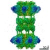
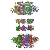
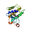
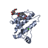
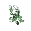
Journal: Proc Natl Acad Sci U S A / Year: 2020 Title: Structure of the cell-binding component of the binary toxin reveals a di-heptamer macromolecular assembly. Authors: Xingjian Xu / Raquel Godoy-Ruiz / Kaylin A Adipietro / Christopher Peralta / Danya Ben-Hail / Kristen M Varney / Mary E Cook / Braden M Roth / Paul T Wilder / Thomas Cleveland / Alexander ...Authors: Xingjian Xu / Raquel Godoy-Ruiz / Kaylin A Adipietro / Christopher Peralta / Danya Ben-Hail / Kristen M Varney / Mary E Cook / Braden M Roth / Paul T Wilder / Thomas Cleveland / Alexander Grishaev / Heather M Neu / Sarah L J Michel / Wenbo Yu / Dorothy Beckett / Richard R Rustandi / Catherine Lancaster / John W Loughney / Adam Kristopeit / Sianny Christanti / Jessica W Olson / Alexander D MacKerell / Amedee des Georges / Edwin Pozharski / David J Weber / Abstract: Targeting infection is challenging because treatment options are limited, and high recurrence rates are common. One reason for this is that hypervirulent strains often have a binary toxin termed ...Targeting infection is challenging because treatment options are limited, and high recurrence rates are common. One reason for this is that hypervirulent strains often have a binary toxin termed the toxin, in addition to the enterotoxins TsdA and TsdB. The toxin has an enzymatic component, termed CDTa, and a pore-forming or delivery subunit termed CDTb. CDTb was characterized here using a combination of single-particle cryoelectron microscopy, X-ray crystallography, NMR, and other biophysical methods. In the absence of CDTa, 2 di-heptamer structures for activated CDTb (1.0 MDa) were solved at atomic resolution, including a symmetric (CDTb; 3.14 Å) and an asymmetric form (CDTb; 2.84 Å). Roles played by 2 receptor-binding domains of activated CDTb were of particular interest since the receptor-binding domain 1 lacks sequence homology to any other known toxin, and the receptor-binding domain 2 is completely absent in other well-studied heptameric toxins (i.e., anthrax). For CDTb, a Ca binding site was discovered in the first receptor-binding domain that is important for its stability, and the second receptor-binding domain was found to be critical for host cell toxicity and the di-heptamer fold for both forms of activated CDTb. Together, these studies represent a starting point for developing structure-based drug-design strategies to target the most severe strains of .
In the structure databanks used in Yorodumi, some data are registered as the other names, "COVID-19 virus" and "2019-nCoV". Here are the details of the virus and the list of structure data.
Jan 31, 2019. EMDB accession codes are about to change! (news from PDBe EMDB page)
EMDB accession codes are about to change! (news from PDBe EMDB page)
The allocation of 4 digits for EMDB accession codes will soon come to an end. Whilst these codes will remain in use, new EMDB accession codes will include an additional digit and will expand incrementally as the available range of codes is exhausted. The current 4-digit format prefixed with “EMD-” (i.e. EMD-XXXX) will advance to a 5-digit format (i.e. EMD-XXXXX), and so on. It is currently estimated that the 4-digit codes will be depleted around Spring 2019, at which point the 5-digit format will come into force.
The EM Navigator/Yorodumi systems omit the EMD- prefix.
Related info.:Q: What is EMD? / ID/Accession-code notation in Yorodumi/EM Navigator
Yorodumi is a browser for structure data from EMDB, PDB, SASBDB, etc.
This page is also the successor to EM Navigator detail page, and also detail information page/front-end page for Omokage search.
The word "yorodu" (or yorozu) is an old Japanese word meaning "ten thousand". "mi" (miru) is to see.
Related info.:EMDB / PDB / SASBDB / Comparison of 3 databanks / Yorodumi Search / Aug 31, 2016. New EM Navigator & Yorodumi / Yorodumi Papers / Jmol/JSmol / Function and homology information / Changes in new EM Navigator and Yorodumi
 Movie
Movie Controller

 Yorodumi
 Open data
 Basic information
 Components
 Keywords
 Function and homology information
 Clostridioides difficile (bacteria)
 X-RAY DIFFRACTION /
  Authors
 Citation
 Structure visualization
 Downloads & links
 Other downloads
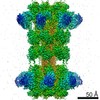


 PDBj
PDBj
 Assembly
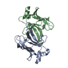

 Mass: 18.015 Da / Num. of mol.: 3 / Source method: isolated from a natural source / Formula: H2O
 Sample preparation
 Processing