 Movie
Movie Controller

[English] 日本語
 Yorodumi
- PDB-6uwt: Clostridium difficile binary toxin translocase CDTb tetradecamer ... -
+ Open data

- Basic information
| Entry | Database: PDB / ID: 6uwt | ||||||
|---|---|---|---|---|---|---|---|
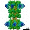
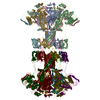
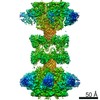
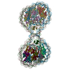
| Title | Clostridium difficile binary toxin translocase CDTb tetradecamer in symmetric conformation | ||||||
 Components Components | ADP-ribosyltransferase binding component | ||||||
 Keywords Keywords | TOXIN / CDTb / translocase / tetradecamer | ||||||
| Function / homology |  Function and homology information Function and homology informationprotein homooligomerization / transferase activity / extracellular region / metal ion binding Similarity search - Function | ||||||
| Biological species |  Clostridioides difficile (bacteria) Clostridioides difficile (bacteria) | ||||||
| Method | ELECTRON MICROSCOPY / single particle reconstruction / cryo EM / Resolution: 3.1 Å | ||||||
 Authors Authors | Xu, X. / Pozharski, E. / des Georges, A. | ||||||
 Citation Citation | Journal: Proc Natl Acad Sci U S A / Year: 2020 Title: Structure of the cell-binding component of the binary toxin reveals a di-heptamer macromolecular assembly. Authors: Xingjian Xu / Raquel Godoy-Ruiz / Kaylin A Adipietro / Christopher Peralta / Danya Ben-Hail / Kristen M Varney / Mary E Cook / Braden M Roth / Paul T Wilder / Thomas Cleveland / Alexander ...Authors: Xingjian Xu / Raquel Godoy-Ruiz / Kaylin A Adipietro / Christopher Peralta / Danya Ben-Hail / Kristen M Varney / Mary E Cook / Braden M Roth / Paul T Wilder / Thomas Cleveland / Alexander Grishaev / Heather M Neu / Sarah L J Michel / Wenbo Yu / Dorothy Beckett / Richard R Rustandi / Catherine Lancaster / John W Loughney / Adam Kristopeit / Sianny Christanti / Jessica W Olson / Alexander D MacKerell / Amedee des Georges / Edwin Pozharski / David J Weber /  Abstract: Targeting infection is challenging because treatment options are limited, and high recurrence rates are common. One reason for this is that hypervirulent strains often have a binary toxin termed ...Targeting infection is challenging because treatment options are limited, and high recurrence rates are common. One reason for this is that hypervirulent strains often have a binary toxin termed the toxin, in addition to the enterotoxins TsdA and TsdB. The toxin has an enzymatic component, termed CDTa, and a pore-forming or delivery subunit termed CDTb. CDTb was characterized here using a combination of single-particle cryoelectron microscopy, X-ray crystallography, NMR, and other biophysical methods. In the absence of CDTa, 2 di-heptamer structures for activated CDTb (1.0 MDa) were solved at atomic resolution, including a symmetric (CDTb; 3.14 Å) and an asymmetric form (CDTb; 2.84 Å). Roles played by 2 receptor-binding domains of activated CDTb were of particular interest since the receptor-binding domain 1 lacks sequence homology to any other known toxin, and the receptor-binding domain 2 is completely absent in other well-studied heptameric toxins (i.e., anthrax). For CDTb, a Ca binding site was discovered in the first receptor-binding domain that is important for its stability, and the second receptor-binding domain was found to be critical for host cell toxicity and the di-heptamer fold for both forms of activated CDTb. Together, these studies represent a starting point for developing structure-based drug-design strategies to target the most severe strains of . | ||||||
| History |
|
- Structure visualization
| Movie |
Movie viewer |
|---|---|
| Structure viewer | Molecule: MolmilJmol/JSmol |
- Downloads & links
-Download
| PDBx/mmCIF format | 6uwt.cif.gz | 1.5 MB | Display | PDBx/mmCIF format |
|---|---|---|---|---|
| PDB format | pdb6uwt.ent.gz | 1.3 MB | Display | PDB format |
| PDBx/mmJSON format | 6uwt.json.gz | Tree view | PDBx/mmJSON format | |
| Others |  Other downloads Other downloads |
-Validation report
| Arichive directory | https://data.pdbj.org/pub/pdb/validation_reports/uw/6uwtftp://data.pdbj.org/pub/pdb/validation_reports/uw/6uwt | HTTPS FTP |
|---|
-Related structure data
| Related structure data | 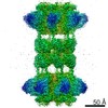 20927MC  6uwiC  6uwoC 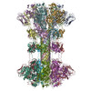 6uwrC M: map data used to model this data C: citing same article ( |
|---|---|
| Similar structure data |
-Links
 PDBj
PDBj
- Assembly
| Deposited unit | 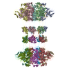
|
|---|---|
| 1 |
|
-Components
| #1: Protein | Mass: 74679.086 Da / Num. of mol.: 14 Source method: isolated from a genetically manipulated source Source: (gene. exp.) Clostridioides difficile (bacteria) / Gene: cdtB / Production host: #2: Chemical | ChemComp-CA /   Mass: 40.078 Da / Num. of mol.: 28 / Source method: obtained synthetically / Formula: Ca Mass: 40.078 Da / Num. of mol.: 28 / Source method: obtained synthetically / Formula: CaHas ligand of interest | N | |
|---|
-Experimental details
-Experiment
| Experiment | Method: ELECTRON MICROSCOPY |
|---|---|
| EM experiment | Aggregation state: PARTICLE / 3D reconstruction method: single particle reconstruction |
- Sample preparation
| Component | Name: CDTb tetradecamer / Type: COMPLEX / Entity ID: #1 / Source: RECOMBINANT |
|---|---|
| Molecular weight | Value: 1.05 MDa / Experimental value: YES |
| Source (natural) | Organism: Clostridioides difficile (bacteria) |
| Source (recombinant) | Organism: |
| Buffer solution | pH: 7.5 |
| Specimen | Embedding applied: NO / Shadowing applied: NO / Staining applied: NO / Vitrification applied: YES |
| Vitrification | Cryogen name: ETHANE |
- Electron microscopy imaging
| Experimental equipment |  Model: Titan Krios / Image courtesy: FEI Company |
|---|---|
| Microscopy | Model: FEI TITAN KRIOS |
| Electron gun | Electron source:  FIELD EMISSION GUN / Accelerating voltage: 300 kV / Illumination mode: SPOT SCAN FIELD EMISSION GUN / Accelerating voltage: 300 kV / Illumination mode: SPOT SCAN |
| Electron lens | Mode: BRIGHT FIELD |
| Image recording | Electron dose: 56.9 e/Å2 / Film or detector model: GATAN K2 SUMMIT (4k x 4k) |
- Processing
| Software |
| ||||||||||||||||||||||||
|---|---|---|---|---|---|---|---|---|---|---|---|---|---|---|---|---|---|---|---|---|---|---|---|---|---|
| CTF correction | Type: PHASE FLIPPING AND AMPLITUDE CORRECTION | ||||||||||||||||||||||||
| Symmetry | Point symmetry: C7 (7 fold cyclic) | ||||||||||||||||||||||||
| 3D reconstruction | Resolution: 3.1 Å / Resolution method: FSC 0.143 CUT-OFF / Num. of particles: 19944 / Symmetry type: POINT | ||||||||||||||||||||||||
| Refinement | Cross valid method: NONE Stereochemistry target values: GeoStd + Monomer Library + CDL v1.2 | ||||||||||||||||||||||||
| Displacement parameters | Biso mean: 21.6 Å2 | ||||||||||||||||||||||||
| Refine LS restraints |
|
