 Movie
Movie Controller

[English] 日本語
 Yorodumi
- PDB-1m8t: Structure of an acidic Phospholipase A2 from the venom of Ophioph... -
+ Open data

- Basic information
| Entry | Database: PDB / ID: 1m8t | ||||||
|---|---|---|---|---|---|---|---|
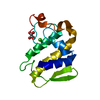
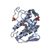
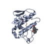
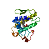
| Title | Structure of an acidic Phospholipase A2 from the venom of Ophiophagus hannah at 2.1 resolution from a hemihedrally twinned crystal form | ||||||
 Components Components | Phospholipase a2 | ||||||
 Keywords Keywords | HYDROLASE / phospholipase a2 structure / twinned crystal / alpha / beta | ||||||
| Function / homology |  Function and homology information Function and homology informationA2-type glycerophospholipase activity / phospholipase A2 / arachidonate secretion / phospholipid metabolic process / lipid catabolic process / phospholipid binding / positive regulation of fibroblast proliferation / fatty acid biosynthetic process / toxin activity / signaling receptor binding ...A2-type glycerophospholipase activity / phospholipase A2 / arachidonate secretion / phospholipid metabolic process / lipid catabolic process / phospholipid binding / positive regulation of fibroblast proliferation / fatty acid biosynthetic process / toxin activity / signaling receptor binding / calcium ion binding / extracellular region Similarity search - Function | ||||||
| Biological species |  Ophiophagus hannah (king cobra) Ophiophagus hannah (king cobra) | ||||||
| Method |  X-RAY DIFFRACTION / MOLECULAR REPLACEMENT / Resolution: 2.1 Å X-RAY DIFFRACTION / MOLECULAR REPLACEMENT / Resolution: 2.1 Å | ||||||
 Authors Authors | Xu, S. / Gu, L. / Wang, Q. / Shu, Y. / Lin, Z. | ||||||
 Citation Citation | |||||||
| History |
|
- Structure visualization
| Structure viewer | Molecule: MolmilJmol/JSmol |
|---|
- Downloads & links
-Download
| PDBx/mmCIF format | 1m8t.cif.gz | 171.2 KB | Display | PDBx/mmCIF format |
|---|---|---|---|---|
| PDB format | pdb1m8t.ent.gz | 135.5 KB | Display | PDB format |
| PDBx/mmJSON format | 1m8t.json.gz | Tree view | PDBx/mmJSON format | |
| Others |  Other downloads Other downloads |
-Validation report
| Arichive directory | https://data.pdbj.org/pub/pdb/validation_reports/m8/1m8tftp://data.pdbj.org/pub/pdb/validation_reports/m8/1m8t | HTTPS FTP |
|---|
-Related structure data
| Related structure data | 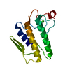 1poaS S: Starting model for refinement |
|---|---|
| Similar structure data |


-Links
 PDBj
PDBj

- Assembly
| Deposited unit | 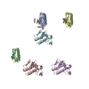
| ||||||||
|---|---|---|---|---|---|---|---|---|---|
| 1 | 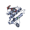
| ||||||||
| 2 | 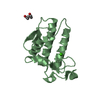
| ||||||||
| 3 | 
| ||||||||
| 4 | 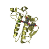
| ||||||||
| 5 | 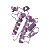
| ||||||||
| 6 | 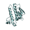
| ||||||||
| Unit cell |
|
-Components
| #1: Protein | Mass: 13200.592 Da / Num. of mol.: 6 / Source method: isolated from a natural source / Source: (natural) Ophiophagus hannah (king cobra) / Secretion: venom / References: UniProt: Q9DF33, phospholipase A2#2: Chemical | ChemComp-CA /   Mass: 40.078 Da / Num. of mol.: 6 / Source method: obtained synthetically / Formula: Ca Mass: 40.078 Da / Num. of mol.: 6 / Source method: obtained synthetically / Formula: Ca#3: Chemical | ChemComp-HEZ /   Mass: 118.174 Da / Num. of mol.: 10 / Source method: obtained synthetically / Formula: C6H14O2 Mass: 118.174 Da / Num. of mol.: 10 / Source method: obtained synthetically / Formula: C6H14O2#4: Water | ChemComp-HOH / |  Mass: 18.015 Da / Num. of mol.: 855 / Source method: isolated from a natural source / Formula: H2O Mass: 18.015 Da / Num. of mol.: 855 / Source method: isolated from a natural source / Formula: H2OHas protein modification | Y | |
|---|
-Experimental details
-Experiment
| Experiment | Method: X-RAY DIFFRACTION / Number of used crystals: 1 |
|---|
- Sample preparation
| Crystal | Density Matthews: 2.32 Å3/Da / Density % sol: 46.96 % | |||||||||||||||||||||||||||||||||||||||||||||||||
|---|---|---|---|---|---|---|---|---|---|---|---|---|---|---|---|---|---|---|---|---|---|---|---|---|---|---|---|---|---|---|---|---|---|---|---|---|---|---|---|---|---|---|---|---|---|---|---|---|---|---|
| Crystal grow | Temperature: 291 K / Method: vapor diffusion, hanging drop / pH: 8.5 Details: 3.4M 1,6-hexanediol, 0.2M magnesium chloride, 0.1M tris-HCl, pH 8.5, VAPOR DIFFUSION, HANGING DROP, temperature 291K | |||||||||||||||||||||||||||||||||||||||||||||||||
| Crystal grow | *PLUS Temperature: 291 K / Method: vapor diffusion, hanging drop | |||||||||||||||||||||||||||||||||||||||||||||||||
| Components of the solutions | *PLUS
|
-Data collection
| Diffraction | Mean temperature: 100 K |
|---|---|
| Diffraction source | Source: ROTATING ANODE / Type: RIGAKU / Wavelength: 1.5418 Å |
| Detector | Type: MARRESEARCH / Detector: AREA DETECTOR / Date: Jan 23, 2002 / Details: Osmic Mirror |
| Radiation | Protocol: SINGLE WAVELENGTH / Monochromatic (M) / Laue (L): M / Scattering type: x-ray |
| Radiation wavelength | Wavelength: 1.5418 Å / Relative weight: 1 |
| Reflection | Resolution: 2.1→17 Å / Num. all: 41966 / Num. obs: 41966 / % possible obs: 99.9 % / Observed criterion σ(I): 0 / Redundancy: 16.1 % / Biso Wilson estimate: 17.7 Å2 / Rmerge(I) obs: 0.078 / Net I/σ(I): 38.1 |
| Reflection shell | Resolution: 2.1→2.17 Å / Redundancy: 14 % / Rmerge(I) obs: 0.383 / Mean I/σ(I) obs: 7.4 / Num. unique all: 4153 / % possible all: 98.8 |
| Reflection | *PLUS Lowest resolution: 17 Å / Num. measured all: 1527037 |
| Reflection shell | *PLUS Highest resolution: 2.1 Å / % possible obs: 98.8 % |
- Processing
| Software |
| ||||||||||||||||||||||||||||||||||||
|---|---|---|---|---|---|---|---|---|---|---|---|---|---|---|---|---|---|---|---|---|---|---|---|---|---|---|---|---|---|---|---|---|---|---|---|---|---|
| Refinement | Method to determine structure: MOLECULAR REPLACEMENT Starting model: PDB ENTRY 1POA Resolution: 2.1→16.99 Å / Rfactor Rfree error: 0.006 / Isotropic thermal model: RESTRAINED / Cross valid method: THROUGHOUT / σ(F): 0 Details: TWINNED R VALUE (WORKING SET): 0.195, TWINNED FREE R VALUE: 0.215; TWINNED BIN R VALUE (WORKING SET): 0.285, TWINNED BIN FREE R VALUE: 0.248
| ||||||||||||||||||||||||||||||||||||
| Solvent computation | Solvent model: FLAT MODEL / Bsol: 95.581 Å2 / ksol: 0.352776 e/Å3 | ||||||||||||||||||||||||||||||||||||
| Displacement parameters | Biso mean: 27.9 Å2
| ||||||||||||||||||||||||||||||||||||
| Refinement step | Cycle: LAST / Resolution: 2.1→16.99 Å
| ||||||||||||||||||||||||||||||||||||
| Refine LS restraints |
| ||||||||||||||||||||||||||||||||||||
| LS refinement shell | Resolution: 2.1→2.23 Å / Total num. of bins used: 6
| ||||||||||||||||||||||||||||||||||||
| Xplor file |
| ||||||||||||||||||||||||||||||||||||
| Refinement | *PLUS Highest resolution: 2.1 Å / Lowest resolution: 17 Å | ||||||||||||||||||||||||||||||||||||
| Solvent computation | *PLUS | ||||||||||||||||||||||||||||||||||||
| Displacement parameters | *PLUS | ||||||||||||||||||||||||||||||||||||
| Refine LS restraints | *PLUS
|
