 Movie
Movie Controller

[English] 日本語
 Yorodumi
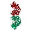
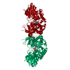
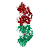
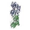
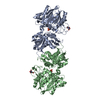
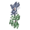
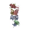
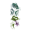
- PDB-6tw0: Human CD73 (ecto 5'-nucleotidase) in complex with PSB12690 (an AO... -
+ Open data

- Basic information
| Entry | Database: PDB / ID: 6tw0 | ||||||
|---|---|---|---|---|---|---|---|
| Title | Human CD73 (ecto 5'-nucleotidase) in complex with PSB12690 (an AOPCP derivative, compound 10 in publication) in the closed state | ||||||
 Components Components | 5'-nucleotidase | ||||||
 Keywords Keywords | HYDROLASE / nucleotide analog / eN / 5NT / complex | ||||||
| Function / homology |  Function and homology information Function and homology informationthymidylate 5'-phosphatase / ADP catabolic process / 5'-deoxynucleotidase / 5'-deoxynucleotidase activity / 7-methylguanosine nucleotidase / adenosine biosynthetic process / inhibition of non-skeletal tissue mineralization / Pyrimidine catabolism / AMP catabolic process / IMP-specific 5'-nucleotidase ...thymidylate 5'-phosphatase / ADP catabolic process / 5'-deoxynucleotidase / 5'-deoxynucleotidase activity / 7-methylguanosine nucleotidase / adenosine biosynthetic process / inhibition of non-skeletal tissue mineralization / Pyrimidine catabolism / AMP catabolic process / IMP-specific 5'-nucleotidase / Purine catabolism / 5'-nucleotidase / 5'-nucleotidase activity / Nicotinate metabolism / leukocyte cell-cell adhesion / DNA metabolic process / response to ATP / Purinergic signaling in leishmaniasis infection / ATP metabolic process / calcium ion homeostasis / negative regulation of inflammatory response / external side of plasma membrane / nucleotide binding / cell surface / extracellular exosome / zinc ion binding / nucleoplasm / membrane / identical protein binding / plasma membrane / cytosol Similarity search - Function | ||||||
| Biological species |  Homo sapiens (human) Homo sapiens (human) | ||||||
| Method |  X-RAY DIFFRACTION / SYNCHROTRON / FOURIER SYNTHESIS / Resolution: 2.5 Å X-RAY DIFFRACTION / SYNCHROTRON / FOURIER SYNTHESIS / Resolution: 2.5 Å | ||||||
 Authors Authors | Pippel, J. / Strater, N. | ||||||
| Funding support |  Germany, 1items Germany, 1items
| ||||||
 Citation Citation | Journal: J.Med.Chem. / Year: 2020 Title: 2-Substituted alpha , beta-Methylene-ADP Derivatives: Potent Competitive Ecto-5'-nucleotidase (CD73) Inhibitors with Variable Binding Modes. Authors: Bhattarai, S. / Pippel, J. / Scaletti, E. / Idris, R. / Freundlieb, M. / Rolshoven, G. / Renn, C. / Lee, S.Y. / Abdelrahman, A. / Zimmermann, H. / El-Tayeb, A. / Muller, C.E. / Strater, N. | ||||||
| History |
|
- Structure visualization
| Structure viewer | Molecule: MolmilJmol/JSmol |
|---|
- Downloads & links
-Download
| PDBx/mmCIF format | 6tw0.cif.gz | 419.1 KB | Display | PDBx/mmCIF format |
|---|---|---|---|---|
| PDB format | pdb6tw0.ent.gz | 345.9 KB | Display | PDB format |
| PDBx/mmJSON format | 6tw0.json.gz | Tree view | PDBx/mmJSON format | |
| Others |  Other downloads Other downloads |
-Validation report
| Arichive directory | https://data.pdbj.org/pub/pdb/validation_reports/tw/6tw0ftp://data.pdbj.org/pub/pdb/validation_reports/tw/6tw0 | HTTPS FTP |
|---|
-Related structure data
| Related structure data |  6tveC  6tvgC  6tvxC  6twaC  6twfC 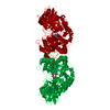 4h2iS S: Starting model for refinement C: citing same article ( |
|---|---|
| Similar structure data |


-Links
 PDBj
PDBj


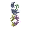
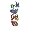
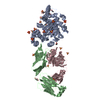
- Assembly
| Deposited unit | 
| ||||||||
|---|---|---|---|---|---|---|---|---|---|
| 1 | 
| ||||||||
| Unit cell |
|
-Components
| #1: Protein | Mass: 59260.406 Da / Num. of mol.: 1 Source method: isolated from a genetically manipulated source Source: (gene. exp.) Homo sapiens (human) / Gene: NT5E, NT5, NTE / Production host:  | ||||||||
|---|---|---|---|---|---|---|---|---|---|
| #2: Chemical |   Mass: 65.409 Da / Num. of mol.: 2 / Source method: obtained synthetically / Formula: Zn Mass: 65.409 Da / Num. of mol.: 2 / Source method: obtained synthetically / Formula: Zn#3: Chemical | ChemComp-NYZ / [[( |   Mass: 441.228 Da / Num. of mol.: 1 / Source method: obtained synthetically / Formula: C11H17N5O10P2 / Feature type: SUBJECT OF INVESTIGATION Mass: 441.228 Da / Num. of mol.: 1 / Source method: obtained synthetically / Formula: C11H17N5O10P2 / Feature type: SUBJECT OF INVESTIGATION#4: Water | ChemComp-HOH / |  Mass: 18.015 Da / Num. of mol.: 15 / Source method: isolated from a natural source / Formula: H2O Mass: 18.015 Da / Num. of mol.: 15 / Source method: isolated from a natural source / Formula: H2OHas ligand of interest | Y | Has protein modification | Y | |
-Experimental details
-Experiment
| Experiment | Method: X-RAY DIFFRACTION / Number of used crystals: 1 |
|---|
- Sample preparation
| Crystal | Density Matthews: 2.64 Å3/Da / Density % sol: 53.39 % |
|---|---|
| Crystal grow | Temperature: 291 K / Method: vapor diffusion, hanging drop Details: Reservoir: 5 % (w/v) polyethylene glycol 6000, 0.1 M NaMES pH 5.5. Drop: 1 microL crystallization buffer + 1 microL of 3 mg/mL protein in 10 mM Tris pH 8.0, 100 microM ZnCl2 and 1 mM of ...Details: Reservoir: 5 % (w/v) polyethylene glycol 6000, 0.1 M NaMES pH 5.5. Drop: 1 microL crystallization buffer + 1 microL of 3 mg/mL protein in 10 mM Tris pH 8.0, 100 microM ZnCl2 and 1 mM of compound. Cryo buffer: 25% PEG200, 0.1 M NaMES pH 5.5 |
-Data collection
| Diffraction | Mean temperature: 100 K / Serial crystal experiment: N | ||||||||||||||||||||||||||||||
|---|---|---|---|---|---|---|---|---|---|---|---|---|---|---|---|---|---|---|---|---|---|---|---|---|---|---|---|---|---|---|---|
| Diffraction source | Source: SYNCHROTRON / Site: BESSY / Beamline: 14.1 / Wavelength: 0.91841 Å | ||||||||||||||||||||||||||||||
| Detector | Type: DECTRIS PILATUS 6M / Detector: PIXEL / Date: Jun 18, 2015 | ||||||||||||||||||||||||||||||
| Radiation | Protocol: SINGLE WAVELENGTH / Monochromatic (M) / Laue (L): M / Scattering type: x-ray | ||||||||||||||||||||||||||||||
| Radiation wavelength | Wavelength: 0.91841 Å / Relative weight: 1 | ||||||||||||||||||||||||||||||
| Reflection | Resolution: 2.5→48.12 Å / Num. obs: 22226 / % possible obs: 99.7 % / Redundancy: 7.3 % / Biso Wilson estimate: 66.87 Å2 / CC1/2: 0.992 / Rmerge(I) obs: 0.349 / Rpim(I) all: 0.137 / Rrim(I) all: 0.375 / Net I/σ(I): 5.2 / Num. measured all: 161402 / Scaling rejects: 2 | ||||||||||||||||||||||||||||||
| Reflection shell | Diffraction-ID: 1
|
- Processing
| Software |
| ||||||||||||||||||||||||||||||||||||||||||||||||||||||||||||||||||||||||||||||||||||||||||||||||||||||||||||
|---|---|---|---|---|---|---|---|---|---|---|---|---|---|---|---|---|---|---|---|---|---|---|---|---|---|---|---|---|---|---|---|---|---|---|---|---|---|---|---|---|---|---|---|---|---|---|---|---|---|---|---|---|---|---|---|---|---|---|---|---|---|---|---|---|---|---|---|---|---|---|---|---|---|---|---|---|---|---|---|---|---|---|---|---|---|---|---|---|---|---|---|---|---|---|---|---|---|---|---|---|---|---|---|---|---|---|---|---|---|
| Refinement | Method to determine structure: FOURIER SYNTHESIS Starting model: 4h2i Resolution: 2.5→48.12 Å / Cor.coef. Fo:Fc: 0.8874 / Cor.coef. Fo:Fc free: 0.8716 / Cross valid method: THROUGHOUT / σ(F): 0 / SU R Blow DPI: 0.479 / SU Rfree Blow DPI: 0.279
| ||||||||||||||||||||||||||||||||||||||||||||||||||||||||||||||||||||||||||||||||||||||||||||||||||||||||||||
| Displacement parameters | Biso max: 202.17 Å2 / Biso mean: 96.91 Å2 / Biso min: 49.6 Å2
| ||||||||||||||||||||||||||||||||||||||||||||||||||||||||||||||||||||||||||||||||||||||||||||||||||||||||||||
| Refine analyze | Luzzati coordinate error obs: 0.633 Å | ||||||||||||||||||||||||||||||||||||||||||||||||||||||||||||||||||||||||||||||||||||||||||||||||||||||||||||
| Refinement step | Cycle: final / Resolution: 2.5→48.12 Å
| ||||||||||||||||||||||||||||||||||||||||||||||||||||||||||||||||||||||||||||||||||||||||||||||||||||||||||||
| Refine LS restraints |
| ||||||||||||||||||||||||||||||||||||||||||||||||||||||||||||||||||||||||||||||||||||||||||||||||||||||||||||
| LS refinement shell | Resolution: 2.5→2.62 Å / Rfactor Rfree error: 0 / Total num. of bins used: 11
| ||||||||||||||||||||||||||||||||||||||||||||||||||||||||||||||||||||||||||||||||||||||||||||||||||||||||||||
| Refinement TLS params. | Method: refined / Refine-ID: X-RAY DIFFRACTION
| ||||||||||||||||||||||||||||||||||||||||||||||||||||||||||||||||||||||||||||||||||||||||||||||||||||||||||||
| Refinement TLS group |
|
