[English] 日本語
Yorodumi
- PDB-6z9b: Human Ecto-5'-nucleotidase (CD73) in complex with AOPCP derivativ... -

+
Open data


ID or keywords:

Loading...

-
Basic information

Entry
Database: PDB / ID: 6z9b
TitleHuman Ecto-5'-nucleotidase (CD73) in complex with AOPCP derivative A830 (compound 16 in publication) in the closed form (crystal form III)
Components5'-nucleotidase
KeywordsHYDROLASE / zinc enzyme / nucleotide analog / inhibitor / Arcus Biosciences
Function / homology
Function and homology information


thymidylate 5'-phosphatase / thymidylate 5'-phosphatase activity / ADP catabolic process / 5'-deoxynucleotidase / 5'-deoxynucleotidase activity / 7-methylguanosine nucleotidase / inhibition of non-skeletal tissue mineralization / adenosine biosynthetic process / Pyrimidine catabolism / GMP 5'-nucleotidase activity ...thymidylate 5'-phosphatase / thymidylate 5'-phosphatase activity / ADP catabolic process / 5'-deoxynucleotidase / 5'-deoxynucleotidase activity / 7-methylguanosine nucleotidase / inhibition of non-skeletal tissue mineralization / adenosine biosynthetic process / Pyrimidine catabolism / GMP 5'-nucleotidase activity / IMP-specific 5'-nucleotidase / IMP 5'-nucleotidase activity / AMP catabolic process / Nicotinate metabolism / Purine catabolism / XMP 5'-nucleosidase activity / 5'-nucleotidase / 5'-nucleotidase activity / leukocyte cell-cell adhesion / DNA metabolic process / response to ATP / : / calcium ion homeostasis / Purinergic signaling in leishmaniasis infection / ATP metabolic process / negative regulation of inflammatory response / external side of plasma membrane / nucleotide binding / cell surface / zinc ion binding / extracellular exosome / nucleoplasm / identical protein binding / membrane / plasma membrane / cytosol
Similarity search - Function
5'-nucleotidase signature 1. / 5'-Nucleotidase, conserved site / 5'-nucleotidase signature 2. / 5'-Nucleotidase, C-terminal / 5'-nucleotidase, C-terminal domain / 5'-Nucleotidase/apyrase / 5'-Nucleotidase, C-terminal domain superfamily / Calcineurin-like phosphoesterase domain, ApaH type / Calcineurin-like phosphoesterase / Metallo-dependent phosphatase-like
Similarity search - Domain/homology
Chem-QCQ / 5'-nucleotidase
Similarity search - Component
Biological speciesHomo sapiens (human)
MethodX-RAY DIFFRACTION / SYNCHROTRON / FOURIER SYNTHESIS / Resolution: 2.17 Å
Model detailsThe compound is a competitive non-nucleotide inhibitor binding to the active site
AuthorsStrater, N. / Scaletti, E.
CitationJournal: J.Med.Chem. / Year: 2020
Title: Discovery of AB680: A Potent and Selective Inhibitor of CD73.
Authors: Lawson, K.V. / Kalisiak, J. / Lindsey, E.A. / Newcomb, E.T. / Leleti, M.R. / Debien, L. / Rosen, B.R. / Miles, D.H. / Sharif, E.U. / Jeffrey, J.L. / Tan, J.B.L. / Chen, A. / Zhao, S. / Xu, G. ...Authors: Lawson, K.V. / Kalisiak, J. / Lindsey, E.A. / Newcomb, E.T. / Leleti, M.R. / Debien, L. / Rosen, B.R. / Miles, D.H. / Sharif, E.U. / Jeffrey, J.L. / Tan, J.B.L. / Chen, A. / Zhao, S. / Xu, G. / Fu, L. / Jin, L. / Park, T.W. / Berry, W. / Moschutz, S. / Scaletti, E. / Strater, N. / Walker, N.P. / Young, S.W. / Walters, M.J. / Schindler, U. / Powers, J.P.
History
DepositionJun 3, 2020Deposition site: PDBE / Processing site: PDBE
Revision 1.0Jul 22, 2020Provider: repository / Type: Initial release
Revision 1.1Jul 29, 2020Group: Database references / Category: citation / citation_author
Item: _citation.title / _citation_author.identifier_ORCID / _citation_author.name
Revision 1.2Nov 4, 2020Group: Database references / Category: citation
Item: _citation.journal_volume / _citation.page_first / _citation.page_last
Revision 1.3Jan 24, 2024Group: Data collection / Database references / Refinement description
Category: chem_comp_atom / chem_comp_bond ...chem_comp_atom / chem_comp_bond / database_2 / pdbx_initial_refinement_model
Item: _database_2.pdbx_DOI / _database_2.pdbx_database_accession

-
Structure visualization

Structure viewerMolecule:
MolmilJmol/JSmol

Downloads & links

-
Assembly

Deposited unit
A: 5'-nucleotidase
hetero molecules


Theoretical massNumber of molelcules
Total (without water)59,9835
Polymers59,2601
Non-polymers7234
Water5,134285
1
A: 5'-nucleotidase
hetero molecules

A: 5'-nucleotidase
hetero molecules


Theoretical massNumber of molelcules
Total (without water)119,96610
Polymers118,5212
Non-polymers1,4458
Water362
TypeNameSymmetry operationNumber
identity operation1_555x,y,z1
crystal symmetry operation3_454-x-1,y,-z-1/21
Buried area2770 Å2
ΔGint-32 kcal/mol
Surface area40190 Å2
Unit cell
Length a, b, c (Å)53.240, 96.450, 235.170
Angle α, β, γ (deg.)90.000, 90.000, 90.000
Int Tables number20
Space group name H-MC2221

-
Components

#1: Protein 5'-nucleotidase / 5'-NT / Ecto-5'-nucleotidase


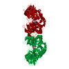
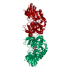
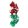
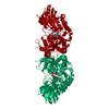
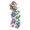
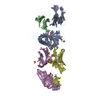
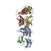
Mass: 59260.406 Da / Num. of mol.: 1
Source method: isolated from a genetically manipulated source
Details: CD73 Construct 4.0 for E. coli expression and refolding
Source: (gene. exp.) Homo sapiens (human) / Gene: NT5E, NT5, NTE / Production host: Escherichia coli (E. coli) / References: UniProt: P21589, 5'-nucleotidase
#2: Chemical ChemComp-ZN / ZINC ION


Mass: 65.409 Da / Num. of mol.: 2 / Source method: obtained synthetically / Formula: Zn
#3: Chemical ChemComp-CA / CALCIUM ION


Mass: 40.078 Da / Num. of mol.: 1 / Source method: obtained synthetically / Formula: Ca
#4: Chemical ChemComp-QCQ / [[(2~{R},3~{R},4~{S},5~{R})-5-[2-chloranyl-6-[(phenylmethyl)amino]purin-9-yl]-4-fluoranyl-3-oxidanyl-oxolan-2-yl]methoxy-oxidanyl-phosphoryl]methylphosphonic acid


Mass: 551.787 Da / Num. of mol.: 1 / Source method: obtained synthetically / Formula: C18H21ClFN5O8P2 / Feature type: SUBJECT OF INVESTIGATION
#5: Water ChemComp-HOH / water


Mass: 18.015 Da / Num. of mol.: 285 / Source method: isolated from a natural source / Formula: H2O
Has ligand of interestY

-
Experimental details

-
Experiment

ExperimentMethod: X-RAY DIFFRACTION / Number of used crystals: 1

-
Sample preparation

CrystalDensity Matthews: 2.55 Å3/Da / Density % sol: 51.71 %
Crystal growTemperature: 291 K / Method: vapor diffusion, hanging drop / pH: 5.5
Details: CD73 at 7 mg/mL was incubated on ice for one hour with 1mM of the inhibitor and 0.1 mM ZnCl2. 1 uL of protein was mixed with 1 uL of 11-14% PEG6000 and 100 mM sodium citrate pH 5.4-5.6. For ...Details: CD73 at 7 mg/mL was incubated on ice for one hour with 1mM of the inhibitor and 0.1 mM ZnCl2. 1 uL of protein was mixed with 1 uL of 11-14% PEG6000 and 100 mM sodium citrate pH 5.4-5.6. For cryoprotection glycerol was added to a final concentration of 15 % to the reservoir buffer.

-
Data collection

DiffractionMean temperature: 100 K / Serial crystal experiment: N
Diffraction sourceSource: SYNCHROTRON / Site: BESSY / Beamline: 14.1 / Wavelength: 0.9184 Å
DetectorType: DECTRIS PILATUS 6M / Detector: PIXEL / Date: Jun 24, 2016
RadiationProtocol: SINGLE WAVELENGTH / Monochromatic (M) / Laue (L): M / Scattering type: x-ray
Radiation wavelengthWavelength: 0.9184 Å / Relative weight: 1
ReflectionResolution: 2.17→48.23 Å / Num. obs: 32496 / % possible obs: 99.2 % / Redundancy: 6.425 % / Biso Wilson estimate: 32.78 Å2 / CC1/2: 0.99 / Rmerge(I) obs: 0.223 / Rrim(I) all: 0.243 / Χ2: 0.803 / Net I/σ(I): 8.61
Reflection shell

Diffraction-ID: 1

Resolution (Å)Redundancy (%)Rmerge(I) obsMean I/σ(I) obsNum. measured obsNum. possibleNum. unique obsCC1/2Rrim(I) all% possible all
2.17-2.36.0380.8361.830625526050720.8370.91596.4
2.3-2.466.8030.6972.5233061487148600.8980.75599.8
2.46-2.656.380.5343.2329120458545640.9380.58299.5
2.65-2.916.6780.3664.9528229423242270.970.39899.9
2.91-3.256.1930.2657.1423615383238130.9810.2999.5
3.25-3.756.5060.18112.9722096340333960.9880.19799.8
3.75-4.586.4870.12920.8818922292129170.990.1499.9
4.58-6.456.4740.10722.4614742228022770.9950.11699.9
6.45-48.236.1210.07228.98319136913590.9980.07999.3

-
Processing

Software
NameVersionClassification
XSCALEdata scaling
BUSTER2.10.3refinement
PDB_EXTRACT3.25data extraction
XDSdata reduction
BUSTERphasing
RefinementMethod to determine structure: FOURIER SYNTHESIS
Starting model: pdbid 4H2I
Resolution: 2.17→48.23 Å / Cor.coef. Fo:Fc: 0.921 / Cor.coef. Fo:Fc free: 0.881 / SU R Cruickshank DPI: 0.245 / Cross valid method: THROUGHOUT / σ(F): 0 / SU R Blow DPI: 0.26 / SU Rfree Blow DPI: 0.213 / SU Rfree Cruickshank DPI: 0.21
RfactorNum. reflection% reflectionSelection details
Rfree0.263 1625 5 %RANDOM
Rwork0.208 ---
obs0.21 32496 99.2 %-
Displacement parametersBiso max: 131.03 Å2 / Biso mean: 42.75 Å2 / Biso min: 18.89 Å2
Baniso -1Baniso -2Baniso -3
1-2.532 Å20 Å20 Å2
2---13.9648 Å20 Å2
3---11.4328 Å2
Refine analyzeLuzzati coordinate error obs: 0.33 Å
Refinement stepCycle: final / Resolution: 2.17→48.23 Å
ProteinNucleic acidLigandSolventTotal
Num. atoms4064 0 38 285 4387
Biso mean--34.52 41.82 -
Num. residues----521
Refine LS restraints
Refine-IDTypeNumberRestraint functionWeightDev ideal
X-RAY DIFFRACTIONt_dihedral_angle_d1464SINUSOIDAL2
X-RAY DIFFRACTIONt_trig_c_planes
X-RAY DIFFRACTIONt_gen_planes730HARMONIC5
X-RAY DIFFRACTIONt_it4198HARMONIC20
X-RAY DIFFRACTIONt_nbd0SEMIHARMONIC5
X-RAY DIFFRACTIONt_improper_torsion
X-RAY DIFFRACTIONt_pseud_angle
X-RAY DIFFRACTIONt_chiral_improper_torsion536SEMIHARMONIC5
X-RAY DIFFRACTIONt_sum_occupancies
X-RAY DIFFRACTIONt_utility_distance
X-RAY DIFFRACTIONt_utility_angle
X-RAY DIFFRACTIONt_utility_torsion
X-RAY DIFFRACTIONt_ideal_dist_contact4960SEMIHARMONIC4
X-RAY DIFFRACTIONt_bond_d4198HARMONIC20.01
X-RAY DIFFRACTIONt_angle_deg5693HARMONIC21.16
X-RAY DIFFRACTIONt_omega_torsion3.48
X-RAY DIFFRACTIONt_other_torsion20.13
LS refinement shellResolution: 2.17→2.18 Å / Rfactor Rfree error: 0 / Total num. of bins used: 50
RfactorNum. reflection% reflection
Rfree0.2175 32 4.92 %
Rwork0.2446 618 -
all0.2434 650 -
obs--86.41 %
Refinement TLS params.

Method: refined / Refine-ID: X-RAY DIFFRACTION

IDL112)L122)L132)L222)L232)L332)S11 (Å °)S12 (Å °)S13 (Å °)S21 (Å °)S22 (Å °)S23 (Å °)S31 (Å °)S32 (Å °)S33 (Å °)T112)T122)T132)T222)T232)T332)Origin x (Å)Origin y (Å)Origin z (Å)
11.0949-0.28470.09440.86720.0032.06520.03830.18170.01470.0332-0.06140.005-0.21270.0380.0231-0.1280.01060.00150.1221-0.0266-0.2224-20.97421.1955-16.2391
20.850.05370.43720.81650.85885.9305-0.13720.12840.00950.03590.2412-0.098-0.12190.5442-0.104-0.29560.00170.02640.2298-0.0151-0.304-17.955218.9849-48.4863
Refinement TLS group
IDRefine-IDRefine TLS-IDSelection detailsAuth asym-IDAuth seq-ID
1X-RAY DIFFRACTION1{A|1 - A|333 A|601 - A|603}A1 - 333
2X-RAY DIFFRACTION1{A|1 - A|333 A|601 - A|603}A601 - 603
3X-RAY DIFFRACTION2{A|334 - A|549}A334 - 549

+
About Yorodumi

-
News

-
Feb 9, 2022. New format data for meta-information of EMDB entries

New format data for meta-information of EMDB entries

  • Version 3 of the EMDB header file is now the official format.
  • The previous official version 1.9 will be removed from the archive.

Related info.:EMDB header

External links:wwPDB to switch to version 3 of the EMDB data model

-
Aug 12, 2020. Covid-19 info

Covid-19 info

URL: https://pdbjlvh1.pdbj.org/emnavi/covid19.php

New page: Covid-19 featured information page in EM Navigator.

Related info.:Covid-19 info / Mar 5, 2020. Novel coronavirus structure data

+
Mar 5, 2020. Novel coronavirus structure data

Novel coronavirus structure data

Related info.:Yorodumi Speices / Aug 12, 2020. Covid-19 info

External links:COVID-19 featured content - PDBj / Molecule of the Month (242):Coronavirus Proteases

+
Jan 31, 2019. EMDB accession codes are about to change! (news from PDBe EMDB page)

EMDB accession codes are about to change! (news from PDBe EMDB page)

  • The allocation of 4 digits for EMDB accession codes will soon come to an end. Whilst these codes will remain in use, new EMDB accession codes will include an additional digit and will expand incrementally as the available range of codes is exhausted. The current 4-digit format prefixed with “EMD-” (i.e. EMD-XXXX) will advance to a 5-digit format (i.e. EMD-XXXXX), and so on. It is currently estimated that the 4-digit codes will be depleted around Spring 2019, at which point the 5-digit format will come into force.
  • The EM Navigator/Yorodumi systems omit the EMD- prefix.

Related info.:Q: What is EMD? / ID/Accession-code notation in Yorodumi/EM Navigator

External links:EMDB Accession Codes are Changing Soon! / Contact to PDBj

+
Jul 12, 2017. Major update of PDB

Major update of PDB

  • wwPDB released updated PDB data conforming to the new PDBx/mmCIF dictionary.
  • This is a major update changing the version number from 4 to 5, and with Remediation, in which all the entries are updated.
  • In this update, many items about electron microscopy experimental information are reorganized (e.g. em_software).
  • Now, EM Navigator and Yorodumi are based on the updated data.

External links:wwPDB Remediation / Enriched Model Files Conforming to OneDep Data Standards Now Available in the PDB FTP Archive

-
Yorodumi

Thousand views of thousand structures

  • Yorodumi is a browser for structure data from EMDB, PDB, SASBDB, etc.
  • This page is also the successor to EM Navigator detail page, and also detail information page/front-end page for Omokage search.
  • The word "yorodu" (or yorozu) is an old Japanese word meaning "ten thousand". "mi" (miru) is to see.

Related info.:EMDB / PDB / SASBDB / Comparison of 3 databanks / Yorodumi Search / Aug 31, 2016. New EM Navigator & Yorodumi / Yorodumi Papers / Jmol/JSmol / Function and homology information / Changes in new EM Navigator and Yorodumi

Read more