 Movie
Movie Controller

[English] 日本語
 Yorodumi
- PDB-6tvg: Human CD73 (ecto 5'-nucleotidase) in complex with AMPCP in the op... -
+ Open data

- Basic information
| Entry | Database: PDB / ID: 6tvg | ||||||
|---|---|---|---|---|---|---|---|
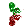
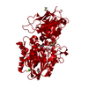
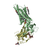
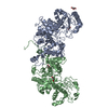
| Title | Human CD73 (ecto 5'-nucleotidase) in complex with AMPCP in the open state | ||||||
 Components Components | 5'-nucleotidase, ecto (CD73), isoform CRA_a | ||||||
 Keywords Keywords | HYDROLASE / eN / e5NT / ecto-nucleotidase / nucleotide | ||||||
| Function / homology |  Function and homology information Function and homology informationthymidylate 5'-phosphatase / ADP catabolic process / 5'-deoxynucleotidase / 5'-deoxynucleotidase activity / 7-methylguanosine nucleotidase / adenosine biosynthetic process / inhibition of non-skeletal tissue mineralization / Pyrimidine catabolism / nucleotide catabolic process / IMP-specific 5'-nucleotidase ...thymidylate 5'-phosphatase / ADP catabolic process / 5'-deoxynucleotidase / 5'-deoxynucleotidase activity / 7-methylguanosine nucleotidase / adenosine biosynthetic process / inhibition of non-skeletal tissue mineralization / Pyrimidine catabolism / nucleotide catabolic process / IMP-specific 5'-nucleotidase / AMP catabolic process / Purine catabolism / 5'-nucleotidase / 5'-nucleotidase activity / Nicotinate metabolism / leukocyte cell-cell adhesion / DNA metabolic process / response to ATP / ATP metabolic process / Purinergic signaling in leishmaniasis infection / calcium ion homeostasis / negative regulation of inflammatory response / external side of plasma membrane / nucleotide binding / cell surface / extracellular exosome / zinc ion binding / nucleoplasm / membrane / metal ion binding / identical protein binding / plasma membrane / cytosol Similarity search - Function | ||||||
| Biological species |  Homo sapiens (human) Homo sapiens (human) | ||||||
| Method |  X-RAY DIFFRACTION / SYNCHROTRON / FOURIER SYNTHESIS / Resolution: 1.48 Å X-RAY DIFFRACTION / SYNCHROTRON / FOURIER SYNTHESIS / Resolution: 1.48 Å | ||||||
 Authors Authors | Scaletti, E. / Strater, N. | ||||||
| Funding support |  Germany, 1items Germany, 1items
| ||||||
 Citation Citation | Journal: J.Med.Chem. / Year: 2020 Title: 2-Substituted alpha , beta-Methylene-ADP Derivatives: Potent Competitive Ecto-5'-nucleotidase (CD73) Inhibitors with Variable Binding Modes. Authors: Bhattarai, S. / Pippel, J. / Scaletti, E. / Idris, R. / Freundlieb, M. / Rolshoven, G. / Renn, C. / Lee, S.Y. / Abdelrahman, A. / Zimmermann, H. / El-Tayeb, A. / Muller, C.E. / Strater, N. | ||||||
| History |
|
- Structure visualization
| Structure viewer | Molecule: MolmilJmol/JSmol |
|---|
- Downloads & links
-Download
| PDBx/mmCIF format | 6tvg.cif.gz | 406.4 KB | Display | PDBx/mmCIF format |
|---|---|---|---|---|
| PDB format | pdb6tvg.ent.gz | 274.5 KB | Display | PDB format |
| PDBx/mmJSON format | 6tvg.json.gz | Tree view | PDBx/mmJSON format | |
| Others |  Other downloads Other downloads |
-Validation report
| Arichive directory | https://data.pdbj.org/pub/pdb/validation_reports/tv/6tvgftp://data.pdbj.org/pub/pdb/validation_reports/tv/6tvg | HTTPS FTP |
|---|
-Related structure data
| Related structure data |  6tveC  6tvxC  6tw0C  6twaC  6twfC  4h2gS S: Starting model for refinement C: citing same article ( |
|---|---|
| Similar structure data |


-Links
 PDBj
PDBj


- Assembly
| Deposited unit | 
| ||||||||||||
|---|---|---|---|---|---|---|---|---|---|---|---|---|---|
| 1 | 
| ||||||||||||
| Unit cell |
| ||||||||||||
| Components on special symmetry positions |
|
-Components
-Protein , 1 types, 1 molecules A
| #1: Protein | Mass: 60464.340 Da / Num. of mol.: 1 Source method: isolated from a genetically manipulated source Source: (gene. exp.) Homo sapiens (human) / Gene: NT5E, hCG_401111 / Production host:  References: UniProt: Q53Z63, UniProt: P21589*PLUS, 5'-nucleotidase |
|---|
-Non-polymers , 5 types, 554 molecules 


| #2: Chemical |  Mass: 65.409 Da / Num. of mol.: 2 / Source method: obtained synthetically / Formula: Zn Mass: 65.409 Da / Num. of mol.: 2 / Source method: obtained synthetically / Formula: Zn#3: Chemical | ChemComp-AP2 / |  Mass: 425.228 Da / Num. of mol.: 1 / Source method: obtained synthetically / Formula: C11H17N5O9P2 / Feature type: SUBJECT OF INVESTIGATION Mass: 425.228 Da / Num. of mol.: 1 / Source method: obtained synthetically / Formula: C11H17N5O9P2 / Feature type: SUBJECT OF INVESTIGATION#4: Chemical | ChemComp-NA / |  Mass: 22.990 Da / Num. of mol.: 1 / Source method: obtained synthetically / Formula: Na Mass: 22.990 Da / Num. of mol.: 1 / Source method: obtained synthetically / Formula: Na#5: Chemical | ChemComp-CA / |  Mass: 40.078 Da / Num. of mol.: 1 / Source method: obtained synthetically / Formula: Ca Mass: 40.078 Da / Num. of mol.: 1 / Source method: obtained synthetically / Formula: Ca#6: Water | ChemComp-HOH / | Mass: 18.015 Da / Num. of mol.: 549 / Source method: isolated from a natural source / Formula: H2O |
|---|
-Details
| Has ligand of interest | Y |
|---|---|
| Has protein modification | Y |
-Experimental details
-Experiment
| Experiment | Method: X-RAY DIFFRACTION / Number of used crystals: 1 |
|---|
- Sample preparation
| Crystal | Density Matthews: 2.42 Å3/Da / Density % sol: 49.11 % |
|---|---|
| Crystal grow | Temperature: 291 K / Method: vapor diffusion, hanging drop Details: 100 mM Tris pH 7.8, 10 % PEG6000, 30 mM AMPCP, 10 % DMSO, 10 % glycerol |
-Data collection
| Diffraction | Mean temperature: 100 K / Serial crystal experiment: N |
|---|---|
| Diffraction source | Source: SYNCHROTRON / Site: BESSY / Beamline: 14.3 / Wavelength: 0.89429 Å |
| Detector | Type: RAYONIX MX-225 / Detector: CCD / Date: Oct 4, 2016 |
| Radiation | Protocol: SINGLE WAVELENGTH / Monochromatic (M) / Laue (L): M / Scattering type: x-ray |
| Radiation wavelength | Wavelength: 0.89429 Å / Relative weight: 1 |
| Reflection | Resolution: 1.477→47.19 Å / Num. obs: 98145 / % possible obs: 99.4 % / Redundancy: 4.96 % / Biso Wilson estimate: 10.7 Å2 / CC1/2: 0.997 / Rrim(I) all: 0.116 / Net I/σ(I): 12 |
| Reflection shell | Resolution: 1.477→1.53 Å / Mean I/σ(I) obs: 1.7 / Num. unique obs: 4759 / CC1/2: 0.554 / Rrim(I) all: 0.993 |
- Processing
| Software |
| |||||||||||||||||||||||||||||||||||||||||||||||||||||||||||||||||||||||||||||||||||||||||||||||||||||||||||||||||||||||||||||||||||||||||||||||||||||||||||||||||||||||||||||||||||||||||||||||||||||||||||||||||||||||||
|---|---|---|---|---|---|---|---|---|---|---|---|---|---|---|---|---|---|---|---|---|---|---|---|---|---|---|---|---|---|---|---|---|---|---|---|---|---|---|---|---|---|---|---|---|---|---|---|---|---|---|---|---|---|---|---|---|---|---|---|---|---|---|---|---|---|---|---|---|---|---|---|---|---|---|---|---|---|---|---|---|---|---|---|---|---|---|---|---|---|---|---|---|---|---|---|---|---|---|---|---|---|---|---|---|---|---|---|---|---|---|---|---|---|---|---|---|---|---|---|---|---|---|---|---|---|---|---|---|---|---|---|---|---|---|---|---|---|---|---|---|---|---|---|---|---|---|---|---|---|---|---|---|---|---|---|---|---|---|---|---|---|---|---|---|---|---|---|---|---|---|---|---|---|---|---|---|---|---|---|---|---|---|---|---|---|---|---|---|---|---|---|---|---|---|---|---|---|---|---|---|---|---|---|---|---|---|---|---|---|---|---|---|---|---|---|---|---|---|
| Refinement | Method to determine structure: FOURIER SYNTHESIS Starting model: 4h2g Resolution: 1.48→47.19 Å / SU ML: 0.1415 / Cross valid method: FREE R-VALUE / σ(F): 1.34 / Phase error: 14.8985 Stereochemistry target values: GeoStd + Monomer Library + CDL v1.2
| |||||||||||||||||||||||||||||||||||||||||||||||||||||||||||||||||||||||||||||||||||||||||||||||||||||||||||||||||||||||||||||||||||||||||||||||||||||||||||||||||||||||||||||||||||||||||||||||||||||||||||||||||||||||||
| Solvent computation | Shrinkage radii: 0.9 Å / VDW probe radii: 1.11 Å / Solvent model: FLAT BULK SOLVENT MODEL | |||||||||||||||||||||||||||||||||||||||||||||||||||||||||||||||||||||||||||||||||||||||||||||||||||||||||||||||||||||||||||||||||||||||||||||||||||||||||||||||||||||||||||||||||||||||||||||||||||||||||||||||||||||||||
| Displacement parameters | Biso mean: 14.9 Å2 | |||||||||||||||||||||||||||||||||||||||||||||||||||||||||||||||||||||||||||||||||||||||||||||||||||||||||||||||||||||||||||||||||||||||||||||||||||||||||||||||||||||||||||||||||||||||||||||||||||||||||||||||||||||||||
| Refinement step | Cycle: LAST / Resolution: 1.48→47.19 Å
| |||||||||||||||||||||||||||||||||||||||||||||||||||||||||||||||||||||||||||||||||||||||||||||||||||||||||||||||||||||||||||||||||||||||||||||||||||||||||||||||||||||||||||||||||||||||||||||||||||||||||||||||||||||||||
| Refine LS restraints |
| |||||||||||||||||||||||||||||||||||||||||||||||||||||||||||||||||||||||||||||||||||||||||||||||||||||||||||||||||||||||||||||||||||||||||||||||||||||||||||||||||||||||||||||||||||||||||||||||||||||||||||||||||||||||||
| LS refinement shell |
|
