 Movie
Movie Controller

[English] 日本語
 Yorodumi
- PDB-6to5: Crystal structure of the oligomerisation domain of the transcript... -
+ Open data

- Basic information
| Entry | Database: PDB / ID: 6to5 | |||||||||
|---|---|---|---|---|---|---|---|---|---|---|
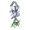
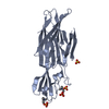
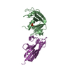
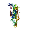
| Title | Crystal structure of the oligomerisation domain of the transcription factor PHOSPHATE STARVATION RESPONSE 1 from Arabidopsis. | |||||||||
 Components Components | Protein PHOSPHATE STARVATION RESPONSE 1 | |||||||||
 Keywords Keywords | TRANSCRIPTION / phosphate starvation / myb domain / coiled-coil domain / inositol pyrophosphate / plant nutrition | |||||||||
| Function / homology |  Function and homology information Function and homology informationresponse to arsenite ion / sulfate ion homeostasis / cellular response to high light intensity / cellular response to phosphate starvation / regulation of monoatomic ion transmembrane transport / circadian rhythm / regulation of gene expression / DNA-binding transcription factor activity / regulation of DNA-templated transcription / DNA binding / nucleus Similarity search - Function | |||||||||
| Biological species |  | |||||||||
| Method |  X-RAY DIFFRACTION / SYNCHROTRON / MOLECULAR REPLACEMENT / Resolution: 2.38 Å X-RAY DIFFRACTION / SYNCHROTRON / MOLECULAR REPLACEMENT / Resolution: 2.38 Å | |||||||||
 Authors Authors | Hothorn, M. | |||||||||
| Funding support |  Switzerland, 2items Switzerland, 2items
| |||||||||
 Citation Citation | Journal: Nat Commun / Year: 2021 Title: Inositol pyrophosphates promote the interaction of SPX domains with the coiled-coil motif of PHR transcription factors to regulate plant phosphate homeostasis. Authors: Ried, M.K. / Wild, R. / Zhu, J. / Pipercevic, J. / Sturm, K. / Broger, L. / Harmel, R.K. / Abriata, L.A. / Hothorn, L.A. / Fiedler, D. / Hiller, S. / Hothorn, M. | |||||||||
| History |
|
- Structure visualization
| Structure viewer | Molecule: MolmilJmol/JSmol |
|---|
- Downloads & links
-Download
| PDBx/mmCIF format | 6to5.cif.gz | 44 KB | Display | PDBx/mmCIF format |
|---|---|---|---|---|
| PDB format | pdb6to5.ent.gz | Display | PDB format | |
| PDBx/mmJSON format | 6to5.json.gz | Tree view | PDBx/mmJSON format | |
| Others |  Other downloads Other downloads |
-Validation report
| Arichive directory | https://data.pdbj.org/pub/pdb/validation_reports/to/6to5ftp://data.pdbj.org/pub/pdb/validation_reports/to/6to5 | HTTPS FTP |
|---|
-Related structure data
| Related structure data |  6to9C  6tocC  5iitS S: Starting model for refinement C: citing same article ( |
|---|---|
| Similar structure data | |
| Experimental dataset #1 | Data reference: 10.5281/zenodo.3570698 / Data set type: diffraction image data / Metadata reference: 10.5281/zenodo.3570698 |

-Links
 PDBj
PDBj
- Assembly
| Deposited unit | 
| ||||||||||||
|---|---|---|---|---|---|---|---|---|---|---|---|---|---|
| 1 | 
| ||||||||||||
| Unit cell |
|
-Components
| #1: Protein | Mass: 9315.647 Da / Num. of mol.: 2 Source method: isolated from a genetically manipulated source Source: (gene. exp.)  #2: Chemical | ChemComp-MG / |   Mass: 24.305 Da / Num. of mol.: 1 / Source method: obtained synthetically / Formula: Mg / Feature type: SUBJECT OF INVESTIGATION Mass: 24.305 Da / Num. of mol.: 1 / Source method: obtained synthetically / Formula: Mg / Feature type: SUBJECT OF INVESTIGATION#3: Water | ChemComp-HOH / |  Mass: 18.015 Da / Num. of mol.: 16 / Source method: isolated from a natural source / Formula: H2O Mass: 18.015 Da / Num. of mol.: 16 / Source method: isolated from a natural source / Formula: H2OHas ligand of interest | Y | |
|---|
-Experimental details
-Experiment
| Experiment | Method: X-RAY DIFFRACTION / Number of used crystals: 1 |
|---|
- Sample preparation
| Crystal | Density Matthews: 2.86 Å3/Da / Density % sol: 56.97 % |
|---|---|
| Crystal grow | Temperature: 298 K / Method: vapor diffusion, sitting drop / pH: 4.2 Details: 0.1 M phosphate citrate pH 4.2, 0.2 M NaCl, 20% (w/v) PEG 8000 |
-Data collection
| Diffraction | Mean temperature: 100 K / Serial crystal experiment: N |
|---|---|
| Diffraction source | Source: SYNCHROTRON / Site: SLS / Beamline: X06DA / Wavelength: 1 Å |
| Detector | Type: DECTRIS PILATUS 2M-F / Detector: PIXEL / Date: Dec 19, 2015 |
| Radiation | Protocol: SINGLE WAVELENGTH / Monochromatic (M) / Laue (L): M / Scattering type: x-ray |
| Radiation wavelength | Wavelength: 1 Å / Relative weight: 1 |
| Reflection | Resolution: 2.38→47.18 Å / Num. obs: 16576 / % possible obs: 99.8 % / Observed criterion σ(I): -3 / Redundancy: 20.8 % / Biso Wilson estimate: 48.58 Å2 / CC1/2: 1 / Rrim(I) all: 0.189 / Net I/σ(I): 14.9 |
| Reflection shell | Resolution: 2.38→2.52 Å / Mean I/σ(I) obs: 1.48 / Num. unique obs: 2262 / CC1/2: 0.69 / Rrim(I) all: 2.21 / % possible all: 98.8 |
- Processing
| Software |
| |||||||||||||||||||||||||||||||||||||||||||||||||
|---|---|---|---|---|---|---|---|---|---|---|---|---|---|---|---|---|---|---|---|---|---|---|---|---|---|---|---|---|---|---|---|---|---|---|---|---|---|---|---|---|---|---|---|---|---|---|---|---|---|---|
| Refinement | Method to determine structure: MOLECULAR REPLACEMENT Starting model: 5IIT Resolution: 2.38→47.17 Å / SU ML: 0.2965 / Cross valid method: FREE R-VALUE / σ(F): 1.34 / Phase error: 25.0838
| |||||||||||||||||||||||||||||||||||||||||||||||||
| Solvent computation | Shrinkage radii: 0.9 Å / VDW probe radii: 1.11 Å | |||||||||||||||||||||||||||||||||||||||||||||||||
| Displacement parameters | Biso mean: 64.43 Å2 | |||||||||||||||||||||||||||||||||||||||||||||||||
| Refinement step | Cycle: LAST / Resolution: 2.38→47.17 Å
| |||||||||||||||||||||||||||||||||||||||||||||||||
| Refine LS restraints |
| |||||||||||||||||||||||||||||||||||||||||||||||||
| LS refinement shell |
|
