 Movie
Movie Controller


+ Open data

- Basic information
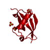
| Entry | Database: PDB / ID: 6sm4 | ||||||
|---|---|---|---|---|---|---|---|
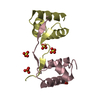
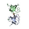
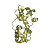
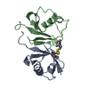
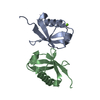
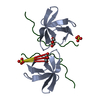
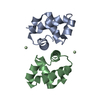
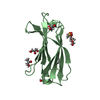
| Title | AntF (apo): type II PKS acyl-carrier protein | ||||||
 Components Components | Acyl carrier protein | ||||||
 Keywords Keywords | PROTEIN BINDING / Natural Product Biosynthesis / Polyketides / Minimal PKS System / Anthraquinone / Chain Elongation / Catalysis | ||||||
| Function / homology | Phosphopantetheine attachment site / ACP-like superfamily / Carrier protein (CP) domain profile. / Phosphopantetheine binding ACP domain / Acyl carrier protein Function and homology information Function and homology information | ||||||
| Biological species |  Photorhabdus luminescens (bacteria) Photorhabdus luminescens (bacteria) | ||||||
| Method |  X-RAY DIFFRACTION / SYNCHROTRON / MOLECULAR REPLACEMENT / Resolution: 1.85 Å X-RAY DIFFRACTION / SYNCHROTRON / MOLECULAR REPLACEMENT / Resolution: 1.85 Å | ||||||
 Authors Authors | Braeuer, A. / Zhou, Q. / Grammbitter, G.L.C. / Schmalhofer, M. / Ruehl, M. / Kaila, V.R.I. / Bode, H. / Groll, M. | ||||||
 Citation Citation | Journal: Nat.Chem. / Year: 2020 Title: Structural snapshots of the minimal PKS system responsible for octaketide biosynthesis. Authors: Brauer, A. / Zhou, Q. / Grammbitter, G.L.C. / Schmalhofer, M. / Ruhl, M. / Kaila, V.R.I. / Bode, H.B. / Groll, M. | ||||||
| History |
|
- Structure visualization
| Structure viewer | Molecule: MolmilJmol/JSmol |
|---|
- Downloads & links
-Download
| PDBx/mmCIF format | 6sm4.cif.gz | 80 KB | Display | PDBx/mmCIF format |
|---|---|---|---|---|
| PDB format | pdb6sm4.ent.gz | 59.4 KB | Display | PDB format |
| PDBx/mmJSON format | 6sm4.json.gz | Tree view | PDBx/mmJSON format | |
| Others |  Other downloads Other downloads |
-Validation report
| Summary document | 6sm4_validation.pdf.gz | 419.6 KB | Display | wwPDB validaton report |
|---|---|---|---|---|
| Full document | 6sm4_full_validation.pdf.gz | 420.3 KB | Display | |
| Data in XML | 6sm4_validation.xml.gz | 8.7 KB | Display | |
| Data in CIF | 6sm4_validation.cif.gz | 11.9 KB | Display | |
| Arichive directory | https://data.pdbj.org/pub/pdb/validation_reports/sm/6sm4ftp://data.pdbj.org/pub/pdb/validation_reports/sm/6sm4 | HTTPS FTP |
-Related structure data
| Related structure data | 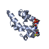 6sm6C  6smdC 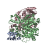 6smoC 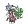 6smpC  2fadS S: Starting model for refinement C: citing same article ( |
|---|---|
| Similar structure data |
-Links
 PDBj
PDBj
- Assembly
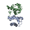
| Deposited unit | 
| ||||||||
|---|---|---|---|---|---|---|---|---|---|
| 1 |
| ||||||||
| Unit cell |
|
-Components
| #1: Protein | Mass: 10850.877 Da / Num. of mol.: 2 Source method: isolated from a genetically manipulated source Source: (gene. exp.) Photorhabdus luminescens (bacteria) / Gene: C6H68_21855 / Production host: #2: Water | ChemComp-HOH / |  Mass: 18.015 Da / Num. of mol.: 132 / Source method: isolated from a natural source / Formula: H2O Mass: 18.015 Da / Num. of mol.: 132 / Source method: isolated from a natural source / Formula: H2O |
|---|
-Experimental details
-Experiment
| Experiment | Method: X-RAY DIFFRACTION / Number of used crystals: 1 |
|---|
- Sample preparation
| Crystal | Density Matthews: 3.7 Å3/Da / Density % sol: 66.73 % |
|---|---|
| Crystal grow | Temperature: 293 K / Method: vapor diffusion, sitting drop / Details: 0.1 M citric acid, 1 M LiCl, 30% PEG 6000 |
-Data collection
| Diffraction | Mean temperature: 100 K / Serial crystal experiment: N |
|---|---|
| Diffraction source | Source: SYNCHROTRON / Site: SLS  / Beamline: X06SA / Wavelength: 1 Å / Beamline: X06SA / Wavelength: 1 Å |
| Detector | Type: DECTRIS EIGER X 16M / Detector: PIXEL / Date: Feb 20, 2016 |
| Radiation | Protocol: SINGLE WAVELENGTH / Monochromatic (M) / Laue (L): M / Scattering type: x-ray |
| Radiation wavelength | Wavelength: 1 Å / Relative weight: 1 |
| Reflection | Resolution: 1.85→50 Å / Num. obs: 23127 / % possible obs: 99.2 % / Redundancy: 7.2 % / CC1/2: 1 / Rmerge(I) obs: 0.043 / Net I/σ(I): 24.5 |
| Reflection shell | Resolution: 1.85→1.95 Å / Rmerge(I) obs: 0.556 / Mean I/σ(I) obs: 3.6 / Num. unique obs: 3301 / CC1/2: 0.923 / % possible all: 99.2 |
- Processing
| Software |
| |||||||||||||||||||||||||||||||||||||||||||||||||||||||||||||||||||||||||||
|---|---|---|---|---|---|---|---|---|---|---|---|---|---|---|---|---|---|---|---|---|---|---|---|---|---|---|---|---|---|---|---|---|---|---|---|---|---|---|---|---|---|---|---|---|---|---|---|---|---|---|---|---|---|---|---|---|---|---|---|---|---|---|---|---|---|---|---|---|---|---|---|---|---|---|---|---|
| Refinement | Method to determine structure: MOLECULAR REPLACEMENT Starting model: 2FAD Resolution: 1.85→15 Å / Cor.coef. Fo:Fc: 0.971 / Cor.coef. Fo:Fc free: 0.956 / SU B: 4.303 / SU ML: 0.058 / Cross valid method: THROUGHOUT / σ(F): 0 / ESU R: 0.111 / ESU R Free: 0.094 Details: HYDROGENS HAVE BEEN ADDED IN THE RIDING POSITIONS U VALUES : REFINED INDIVIDUALLY
| |||||||||||||||||||||||||||||||||||||||||||||||||||||||||||||||||||||||||||
| Solvent computation | Ion probe radii: 0.8 Å / Shrinkage radii: 0.8 Å / VDW probe radii: 1.2 Å | |||||||||||||||||||||||||||||||||||||||||||||||||||||||||||||||||||||||||||
| Displacement parameters | Biso max: 103.68 Å2 / Biso mean: 37.186 Å2 / Biso min: 20.82 Å2
| |||||||||||||||||||||||||||||||||||||||||||||||||||||||||||||||||||||||||||
| Refinement step | Cycle: final / Resolution: 1.85→15 Å
| |||||||||||||||||||||||||||||||||||||||||||||||||||||||||||||||||||||||||||
| Refine LS restraints |
| |||||||||||||||||||||||||||||||||||||||||||||||||||||||||||||||||||||||||||
| LS refinement shell | Resolution: 1.85→1.897 Å / Rfactor Rfree error: 0 / Total num. of bins used: 20
|
