 Movie
Movie Controller


+ Open data

- Basic information
| Entry | Database: PDB / ID: 2fad | ||||||
|---|---|---|---|---|---|---|---|
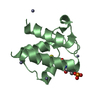
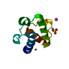
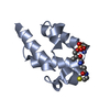
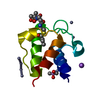
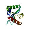
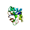
| Title | Crystal structure of E. coli heptanoyl-ACP | ||||||
 Components Components | Acyl carrier protein | ||||||
 Keywords Keywords | BIOSYNTHETIC PROTEIN / acyl carrier protein / acyl chain binding / fatty acid biosynthesis | ||||||
| Function / homology |  Function and homology information Function and homology informationlipid A biosynthetic process / lipid biosynthetic process / phosphopantetheine binding / acyl binding / acyl carrier activity / fatty acid biosynthetic process / response to xenobiotic stimulus / lipid binding / membrane / cytoplasm / cytosol Similarity search - Function | ||||||
| Biological species |  | ||||||
| Method |  X-RAY DIFFRACTION / SYNCHROTRON / MOLECULAR REPLACEMENT / Resolution: 1.6 Å X-RAY DIFFRACTION / SYNCHROTRON / MOLECULAR REPLACEMENT / Resolution: 1.6 Å | ||||||
 Authors Authors | Roujeinikova, A. | ||||||
 Citation Citation | Journal: J.Mol.Biol. / Year: 2007 Title: Structural Studies of Fatty Acyl-(Acyl Carrier Protein) Thioesters Reveal a Hydrophobic Binding Cavity that Can Expand to Fit Longer Substrates. Authors: Roujeinikova, A. / Simon, W.J. / Gilroy, J. / Rice, D.W. / Rafferty, J.B. / Slabas, A.R. | ||||||
| History |
|
- Structure visualization
| Structure viewer | Molecule: MolmilJmol/JSmol |
|---|
- Downloads & links
-Download
| PDBx/mmCIF format | 2fad.cif.gz | 49.7 KB | Display | PDBx/mmCIF format |
|---|---|---|---|---|
| PDB format | pdb2fad.ent.gz | 35.4 KB | Display | PDB format |
| PDBx/mmJSON format | 2fad.json.gz | Tree view | PDBx/mmJSON format | |
| Others |  Other downloads Other downloads |
-Validation report
| Arichive directory | https://data.pdbj.org/pub/pdb/validation_reports/fa/2fadftp://data.pdbj.org/pub/pdb/validation_reports/fa/2fad | HTTPS FTP |
|---|
-Related structure data
| Related structure data |  2facC  2faeC  1l0iS S: Starting model for refinement C: citing same article ( |
|---|---|
| Similar structure data |


-Links
 PDBj
PDBj
- Assembly
| Deposited unit | 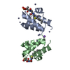
| ||||||||||||||||||
|---|---|---|---|---|---|---|---|---|---|---|---|---|---|---|---|---|---|---|---|
| 1 | 
| ||||||||||||||||||
| 2 | 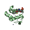
| ||||||||||||||||||
| 3 | 
| ||||||||||||||||||
| Unit cell |
| ||||||||||||||||||
| Components on special symmetry positions |
|
-Components
| #1: Protein | Mass: 8514.264 Da / Num. of mol.: 2 Source method: isolated from a genetically manipulated source Source: (gene. exp.) #2: Chemical | ChemComp-NA / |   Mass: 22.990 Da / Num. of mol.: 1 / Source method: obtained synthetically / Formula: Na Mass: 22.990 Da / Num. of mol.: 1 / Source method: obtained synthetically / Formula: Na#3: Chemical | ChemComp-ZN /   Mass: 65.409 Da / Num. of mol.: 8 / Source method: obtained synthetically / Formula: Zn Mass: 65.409 Da / Num. of mol.: 8 / Source method: obtained synthetically / Formula: Zn#4: Chemical |   Mass: 454.518 Da / Num. of mol.: 2 / Source method: obtained synthetically / Formula: C18H35N2O7PS Mass: 454.518 Da / Num. of mol.: 2 / Source method: obtained synthetically / Formula: C18H35N2O7PS#5: Water | ChemComp-HOH / |  Mass: 18.015 Da / Num. of mol.: 191 / Source method: isolated from a natural source / Formula: H2O Mass: 18.015 Da / Num. of mol.: 191 / Source method: isolated from a natural source / Formula: H2OHas protein modification | Y | |
|---|
-Experimental details
-Experiment
| Experiment | Method: X-RAY DIFFRACTION / Number of used crystals: 1 |
|---|
- Sample preparation
| Crystal | Density Matthews: 2.17 Å3/Da / Density % sol: 43.21 % |
|---|---|
| Crystal grow | Temperature: 290 K / Method: vapor diffusion, hanging drop / pH: 6 Details: 8-12% PEG 4000, 30 mM zinc acetate, 50 mM sodium cocadylate, pH 6.0, VAPOR DIFFUSION, HANGING DROP, temperature 290K |
-Data collection
| Diffraction | Mean temperature: 100 K |
|---|---|
| Diffraction source | Source: SYNCHROTRON / Site: ESRF  / Beamline: ID14-2 / Wavelength: 0.933 Å / Beamline: ID14-2 / Wavelength: 0.933 Å |
| Radiation | Protocol: SINGLE WAVELENGTH / Monochromatic (M) / Laue (L): M / Scattering type: x-ray |
| Radiation wavelength | Wavelength: 0.933 Å / Relative weight: 1 |
| Reflection | Resolution: 1.6→15 Å / Num. obs: 16351 / % possible obs: 80 % / Observed criterion σ(F): 0 / Observed criterion σ(I): 0 / Redundancy: 2.7 % / Biso Wilson estimate: 16 Å2 / Rmerge(I) obs: 0.056 / Net I/σ(I): 19.3 |
| Reflection shell | Resolution: 1.6→1.66 Å / Rmerge(I) obs: 0.4 / Mean I/σ(I) obs: 2.2 / Num. unique all: 1477 / % possible all: 74 |
- Processing
| Software |
| ||||||||||||||||||||||||
|---|---|---|---|---|---|---|---|---|---|---|---|---|---|---|---|---|---|---|---|---|---|---|---|---|---|
| Refinement | Method to determine structure: MOLECULAR REPLACEMENT Starting model: PDB 1L0I Resolution: 1.6→10 Å / σ(F): 0
| ||||||||||||||||||||||||
| Solvent computation | Bsol: 60.276 Å2 | ||||||||||||||||||||||||
| Displacement parameters | Biso mean: 21.289 Å2
| ||||||||||||||||||||||||
| Refinement step | Cycle: LAST / Resolution: 1.6→10 Å
| ||||||||||||||||||||||||
| Refine LS restraints |
| ||||||||||||||||||||||||
| Xplor file |
|
