 Movie
Movie Controller


+ Open data

- Basic information
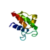
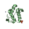
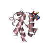
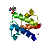
| Entry | Database: PDB / ID: 1l0h | ||||||
|---|---|---|---|---|---|---|---|
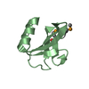
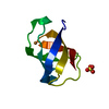
| Title | CRYSTAL STRUCTURE OF BUTYRYL-ACP FROM E.COLI | ||||||
 Components Components | ACYL CARRIER PROTEIN | ||||||
 Keywords Keywords | LIPID TRANSPORT / acyl carrier protein / acyl chain binding / fatty acid biosynthesis | ||||||
| Function / homology |  Function and homology information Function and homology informationlipid A biosynthetic process / lipid biosynthetic process / phosphopantetheine binding / acyl binding / acyl carrier activity / fatty acid biosynthetic process / response to xenobiotic stimulus / lipid binding / membrane / cytoplasm / cytosol Similarity search - Function | ||||||
| Biological species |  | ||||||
| Method |  X-RAY DIFFRACTION / SYNCHROTRON / MOLECULAR REPLACEMENT / Resolution: 2 Å X-RAY DIFFRACTION / SYNCHROTRON / MOLECULAR REPLACEMENT / Resolution: 2 Å | ||||||
 Authors Authors | Roujeinikova, A. / Baldock, C. / Simon, W.J. / Gilroy, J. / Baker, P.J. / Stuitje, A.R. / Rice, D.W. / Slabas, A.R. / Rafferty, J.B. | ||||||
 Citation Citation | Journal: Structure / Year: 2002 Title: X-ray crystallographic studies on butyryl-ACP reveal flexibility of the structure around a putative acyl chain binding site Authors: Roujeinikova, A. / Baldock, C. / Simon, W.J. / Gilroy, J. / Baker, P.J. / Stuitje, A.R. / Rice, D.W. / Slabas, A.R. / Rafferty, J.B. #1: Journal: Acta Crystallogr.,Sect.D / Year: 2002Title: Crystallisation and preliminary X-ray crystallographic studies on acyl-(acyl carrier protein) from Escherichia coli Authors: Roujeinikova, A. / Baldock, C. / Simon, W.J. / Gilroy, J. / Baker, P.J. / Stuitje, A.R. / Rice, D.W. / Rafferty, J.B. / Slabas, A.R. | ||||||
| History |
|
- Structure visualization
| Structure viewer | Molecule: MolmilJmol/JSmol |
|---|
- Downloads & links
-Download
| PDBx/mmCIF format | 1l0h.cif.gz | 28.5 KB | Display | PDBx/mmCIF format |
|---|---|---|---|---|
| PDB format | pdb1l0h.ent.gz | 18.1 KB | Display | PDB format |
| PDBx/mmJSON format | 1l0h.json.gz | Tree view | PDBx/mmJSON format | |
| Others |  Other downloads Other downloads |
-Validation report
| Arichive directory | https://data.pdbj.org/pub/pdb/validation_reports/l0/1l0hftp://data.pdbj.org/pub/pdb/validation_reports/l0/1l0h | HTTPS FTP |
|---|
-Related structure data
| Related structure data |  1l0iSC S: Starting model for refinement C: citing same article ( |
|---|---|
| Similar structure data |


-Links
 PDBj
PDBj
- Assembly
| Deposited unit | 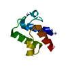
| ||||||||
|---|---|---|---|---|---|---|---|---|---|
| 1 |
| ||||||||
| Unit cell |
|
-Components
| #1: Protein | Mass: 8645.460 Da / Num. of mol.: 1 Source method: isolated from a genetically manipulated source Source: (gene. exp.) | ||
|---|---|---|---|
| #2: Chemical |   Mass: 65.409 Da / Num. of mol.: 2 / Source method: obtained synthetically / Formula: Zn Mass: 65.409 Da / Num. of mol.: 2 / Source method: obtained synthetically / Formula: Zn#3: Water | ChemComp-HOH / |  Mass: 18.015 Da / Num. of mol.: 58 / Source method: isolated from a natural source / Formula: H2O Mass: 18.015 Da / Num. of mol.: 58 / Source method: isolated from a natural source / Formula: H2O |
-Experimental details
-Experiment
| Experiment | Method: X-RAY DIFFRACTION / Number of used crystals: 1 |
|---|
- Sample preparation
| Crystal | Density Matthews: 2.48 Å3/Da / Density % sol: 50.46 % | |||||||||||||||||||||||||||||||||||
|---|---|---|---|---|---|---|---|---|---|---|---|---|---|---|---|---|---|---|---|---|---|---|---|---|---|---|---|---|---|---|---|---|---|---|---|---|
| Crystal grow | Temperature: 290 K / Method: vapor diffusion, hanging drop / pH: 6 Details: PEG 20000, zinc chloride, sodium cacodylate, pH 6.0, VAPOR DIFFUSION, HANGING DROP, temperature 290K | |||||||||||||||||||||||||||||||||||
| Crystal grow | *PLUS Details: Roujeinikova, A., (2002) Acta Crystallogr., Sect.D, 58, 330. | |||||||||||||||||||||||||||||||||||
| Components of the solutions | *PLUS
|
-Data collection
| Diffraction | Mean temperature: 100 K |
|---|---|
| Diffraction source | Source: SYNCHROTRON / Site: ESRF  / Beamline: BM30A / Wavelength: 0.97 Å / Beamline: BM30A / Wavelength: 0.97 Å |
| Detector | Detector: CCD |
| Radiation | Protocol: SINGLE WAVELENGTH / Monochromatic (M) / Laue (L): M / Scattering type: x-ray |
| Radiation wavelength | Wavelength: 0.97 Å / Relative weight: 1 |
| Reflection | Resolution: 2→20 Å / Num. obs: 10616 / Observed criterion σ(F): 0 / Observed criterion σ(I): 0 / Rmerge(I) obs: 0.06 |
| Reflection shell | Resolution: 2→2.05 Å / Rmerge(I) obs: 0.269 |
| Reflection | *PLUS Num. obs: 5485 / % possible obs: 94 % / Num. measured all: 29701 / Rmerge(I) obs: 0.06 |
| Reflection shell | *PLUS % possible obs: 90 % |
- Processing
| Software |
| |||||||||||||||||||||||||
|---|---|---|---|---|---|---|---|---|---|---|---|---|---|---|---|---|---|---|---|---|---|---|---|---|---|---|
| Refinement | Method to determine structure: MOLECULAR REPLACEMENT Starting model: ACP I62M mutant, PDB ENTRY 1L0I Resolution: 2→10 Å / Cross valid method: THROUGHOUT / σ(F): 0 / σ(I): 0
| |||||||||||||||||||||||||
| Displacement parameters | Biso mean: 36 Å2 | |||||||||||||||||||||||||
| Refinement step | Cycle: LAST / Resolution: 2→10 Å
| |||||||||||||||||||||||||
| Refine LS restraints |
| |||||||||||||||||||||||||
| Refinement | *PLUS Lowest resolution: 10 Å / Num. reflection obs: 9273 / % reflection Rfree: 5 % | |||||||||||||||||||||||||
| Solvent computation | *PLUS | |||||||||||||||||||||||||
| Displacement parameters | *PLUS |
