METAL TRANSPORT / CATION PI / METAL-BINDING / COPPER TOLERANCE / COPPER TRANSPORT
Function / homology
Function and homology information
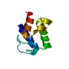
metallochaperone activity / copper ion transmembrane transport / response to silver ion / silver ion transmembrane transport / copper chaperone activity / copper ion export / detoxification of copper ion / response to zinc ion / response to copper ion / transition metal ion binding ...metallochaperone activity / copper ion transmembrane transport / response to silver ion / silver ion transmembrane transport / copper chaperone activity / copper ion export / detoxification of copper ion / response to zinc ion / response to copper ion / transition metal ion binding / intracellular copper ion homeostasis / response to toxic substance / outer membrane-bounded periplasmic space / periplasmic space / copper ion binding Similarity search - Function
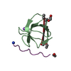
Copper binding periplasmic protein CusF / Copper binding periplasmic protein CusF / Copper binding periplasmic protein CusF / Copper binding periplasmic protein CusF superfamily / OB fold (Dihydrolipoamide Acetyltransferase, E2P) / Beta Barrel / Mainly Beta Similarity search - Domain/homology
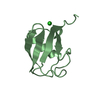
SHEET DETERMINATION METHOD: DSSP THE SHEETS PRESENTED AS "XA" IN EACH CHAIN ON SHEET RECORDS BELOW ... SHEET DETERMINATION METHOD: DSSP THE SHEETS PRESENTED AS "XA" IN EACH CHAIN ON SHEET RECORDS BELOW IS ACTUALLY AN 7-STRANDED BARREL THIS IS REPRESENTED BY A 8-STRANDED SHEET IN WHICH THE FIRST AND LAST STRANDS ARE IDENTICAL.
 Movie
Movie Controller


 Open data
 Basic information
 Components
 Keywords
 Function and homology information
 X-RAY DIFFRACTION /
  Authors
 Citation
 Structure visualization
 Downloads & links
 Other downloads


 PDBj
PDBj Assembly
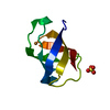

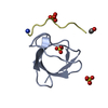
 Mass: 96.063 Da / Num. of mol.: 1 / Source method: obtained synthetically / Formula: SO4
 Mass: 63.546 Da / Num. of mol.: 1 / Source method: obtained synthetically / Formula: Cu
 Mass: 18.015 Da / Num. of mol.: 46 / Source method: isolated from a natural source / Formula: H2O
 Sample preparation
 / Beamline: 17-ID / Wavelength: 1
  Processing