[English] 日本語
Yorodumi
- PDB-4a20: Crystal structure of the Ubl domain of Mdy2 (Get5) at 1.78A -

+
Open data


ID or keywords:

Loading...

-
Basic information

Entry
Database: PDB / ID: 4a20
TitleCrystal structure of the Ubl domain of Mdy2 (Get5) at 1.78A
ComponentsUBIQUITIN-LIKE PROTEIN MDY2
KeywordsPROTEIN BINDING / GET-PATHWAY / TAIL-ANCHORED PROTEINS
Function / homology
Function and homology information


cell morphogenesis involved in conjugation with cellular fusion / TRC complex / tail-anchored membrane protein insertion into ER membrane / protein insertion into ER membrane / post-translational protein targeting to endoplasmic reticulum membrane / cytoplasmic stress granule / protein-folding chaperone binding / nucleus / cytoplasm / cytosol
Similarity search - Function
Mdy2, Get4 binding domain / Get5, C-terminal domain / Binding domain to Get4 on Get5, Golgi to ER traffic protein / Ubiquitin-like protein MDY2, C-terminal domain / Phosphatidylinositol 3-kinase Catalytic Subunit; Chain A, domain 1 / Ubiquitin-like (UB roll) / Ubiquitin family / Ubiquitin homologues / Ubiquitin domain profile. / Ubiquitin-like domain ...Mdy2, Get4 binding domain / Get5, C-terminal domain / Binding domain to Get4 on Get5, Golgi to ER traffic protein / Ubiquitin-like protein MDY2, C-terminal domain / Phosphatidylinositol 3-kinase Catalytic Subunit; Chain A, domain 1 / Ubiquitin-like (UB roll) / Ubiquitin family / Ubiquitin homologues / Ubiquitin domain profile. / Ubiquitin-like domain / Ubiquitin-like domain superfamily / Roll / Alpha Beta
Similarity search - Domain/homology
Ubiquitin-like protein MDY2
Similarity search - Component
Biological speciesSACCHAROMYCES CEREVISIAE (brewer's yeast)
MethodX-RAY DIFFRACTION / SYNCHROTRON / MOLECULAR REPLACEMENT / Resolution: 1.78 Å
AuthorsSimon, A.C. / Simpson, P.J. / Murray, J.W. / Isaacson, R.L.
CitationJournal: Proc.Natl.Acad.Sci.USA / Year: 2013
Title: Structure of the Sgt2/Get5 Complex Provides Insights Into Get-Mediated Targeting of Tail-Anchored Membrane Proteins
Authors: Simon, A.C. / Simpson, P.J. / Goldstone, R.M. / Krysztofinska, E.M. / Murray, J.W. / High, S. / Isaacson, R.L.
History
DepositionSep 21, 2011Deposition site: PDBE / Processing site: PDBE
Revision 1.0Nov 14, 2012Provider: repository / Type: Initial release
Revision 1.1Jan 16, 2013Group: Database references
Revision 1.2Jan 30, 2013Group: Database references
Revision 1.3Feb 13, 2013Group: Database references
Revision 1.4May 8, 2019Group: Data collection / Experimental preparation / Other
Category: database_PDB_rev / database_PDB_rev_record ...database_PDB_rev / database_PDB_rev_record / exptl_crystal_grow / pdbx_database_proc / pdbx_database_status / pdbx_seq_map_depositor_info
Item: _exptl_crystal_grow.method / _pdbx_database_status.recvd_author_approval ..._exptl_crystal_grow.method / _pdbx_database_status.recvd_author_approval / _pdbx_seq_map_depositor_info.one_letter_code / _pdbx_seq_map_depositor_info.one_letter_code_mod
Revision 1.5Dec 20, 2023Group: Data collection / Database references ...Data collection / Database references / Derived calculations / Other / Refinement description
Category: chem_comp_atom / chem_comp_bond ...chem_comp_atom / chem_comp_bond / database_2 / pdbx_database_status / pdbx_initial_refinement_model / struct_site
Item: _database_2.pdbx_DOI / _database_2.pdbx_database_accession ..._database_2.pdbx_DOI / _database_2.pdbx_database_accession / _pdbx_database_status.status_code_sf / _struct_site.pdbx_auth_asym_id / _struct_site.pdbx_auth_comp_id / _struct_site.pdbx_auth_seq_id

-
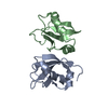
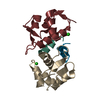
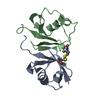
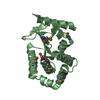
Structure visualization

Structure viewerMolecule:
MolmilJmol/JSmol

Downloads & links

-
Assembly

Deposited unit
A: UBIQUITIN-LIKE PROTEIN MDY2
hetero molecules


Theoretical massNumber of molelcules
Total (without water)11,4054
Polymers11,1171
Non-polymers2883
Water82946
1
A: UBIQUITIN-LIKE PROTEIN MDY2
hetero molecules

A: UBIQUITIN-LIKE PROTEIN MDY2
hetero molecules


Theoretical massNumber of molelcules
Total (without water)22,8108
Polymers22,2342
Non-polymers5766
Water362
TypeNameSymmetry operationNumber
identity operation1_555x,y,z1
crystal symmetry operation5_555x-y,-y,-z+1/31
Buried area2420 Å2
ΔGint-79.1 kcal/mol
Surface area8630 Å2
MethodPISA
Unit cell
Length a, b, c (Å)47.190, 47.190, 78.410
Angle α, β, γ (deg.)90.00, 90.00, 120.00
Int Tables number154
Space group name H-MP3221

-
Components

#1: Protein UBIQUITIN-LIKE PROTEIN MDY2 / GOLGI TO ER TRAFFIC PROTEIN 5 / MATING-DEFICIENT PROTEIN 2 / TRANSLATION MACHINERY-ASSOCIATED PROTEIN 24


Mass: 11116.803 Da / Num. of mol.: 1 / Fragment: GET5 UBL-LIKE DOMAIN, RESIDUES 70-152
Source method: isolated from a genetically manipulated source
Source: (gene. exp.) SACCHAROMYCES CEREVISIAE (brewer's yeast)
Production host: ESCHERICHIA COLI (E. coli) / Strain (production host): BL21(DE3) / References: UniProt: Q12285
#2: Chemical ChemComp-SO4 / SULFATE ION


Mass: 96.063 Da / Num. of mol.: 3 / Source method: obtained synthetically / Formula: SO4
#3: Water ChemComp-HOH / water


Mass: 18.015 Da / Num. of mol.: 46 / Source method: isolated from a natural source / Formula: H2O

-
Experimental details

-
Experiment

ExperimentMethod: X-RAY DIFFRACTION / Number of used crystals: 1

-
Sample preparation

CrystalDensity Matthews: 2.83 Å3/Da / Density % sol: 57 % / Description: NONE
Crystal growMethod: vapor diffusion, hanging drop / pH: 5.5
Details: 3 M AMMONIUM SULFATE, 100 MM SODIUM CITRATE PH 5.5 MIXED WITH AN EQUAL VOLUME OF PROTEIN AT 6MG/ML, HANGING DROP VAPOUR DIFFUSION.

-
Data collection

DiffractionMean temperature: 100 K
Diffraction sourceSource: SYNCHROTRON / Site: Diamond / Beamline: I02 / Wavelength: 0.9795
DetectorType: ADSC CCD / Detector: CCD / Date: Aug 1, 2011
RadiationProtocol: SINGLE WAVELENGTH / Monochromatic (M) / Laue (L): M / Scattering type: x-ray
Radiation wavelengthWavelength: 0.9795 Å / Relative weight: 1
ReflectionResolution: 1.78→40.87 Å / Num. obs: 10128 / % possible obs: 99.8 % / Observed criterion σ(I): 0 / Redundancy: 7.72 % / Rmerge(I) obs: 0.06 / Net I/σ(I): 5.75
Reflection shellResolution: 1.78→1.88 Å / Redundancy: 7.8 % / Rmerge(I) obs: 0.35 / Mean I/σ(I) obs: 2.2 / % possible all: 100

-
Processing

Software
NameVersionClassification
REFMAC5.5.0109refinement
MOSFLMdata reduction
SCALAdata scaling
PHASERphasing
RefinementMethod to determine structure: MOLECULAR REPLACEMENT
Starting model: PDB ENTRY 3M62
Resolution: 1.78→19.77 Å / Cor.coef. Fo:Fc: 0.955 / Cor.coef. Fo:Fc free: 0.93 / SU B: 5.391 / SU ML: 0.077 / Cross valid method: THROUGHOUT / ESU R: 0.116 / ESU R Free: 0.12 / Stereochemistry target values: MAXIMUM LIKELIHOOD / Details: HYDROGENS HAVE BEEN ADDED IN THE RIDING POSITIONS.
RfactorNum. reflection% reflectionSelection details
Rfree0.24825 486 4.8 %RANDOM
Rwork0.20113 ---
obs0.20327 9603 99.72 %-
Solvent computationIon probe radii: 0.8 Å / Shrinkage radii: 0.8 Å / VDW probe radii: 1.4 Å / Solvent model: MASK
Displacement parametersBiso mean: 24.108 Å2
Baniso -1Baniso -2Baniso -3
1-0.01 Å20 Å20 Å2
2--0.01 Å20 Å2
3----0.01 Å2
Refinement stepCycle: LAST / Resolution: 1.78→19.77 Å
ProteinNucleic acidLigandSolventTotal
Num. atoms622 0 15 46 683
Refine LS restraints
Refine-IDTypeDev idealDev ideal targetNumber
X-RAY DIFFRACTIONr_bond_refined_d0.0240.022651
X-RAY DIFFRACTIONr_bond_other_d0.0010.02423
X-RAY DIFFRACTIONr_angle_refined_deg1.9871.997885
X-RAY DIFFRACTIONr_angle_other_deg0.96831063
X-RAY DIFFRACTIONr_dihedral_angle_1_deg5.925581
X-RAY DIFFRACTIONr_dihedral_angle_2_deg39.85126.52223
X-RAY DIFFRACTIONr_dihedral_angle_3_deg14.31515124
X-RAY DIFFRACTIONr_dihedral_angle_4_deg
X-RAY DIFFRACTIONr_chiral_restr0.1140.2109
X-RAY DIFFRACTIONr_gen_planes_refined0.0090.021675
X-RAY DIFFRACTIONr_gen_planes_other0.0010.02101
X-RAY DIFFRACTIONr_nbd_refined0.2210.298
X-RAY DIFFRACTIONr_nbd_other0.1840.2407
X-RAY DIFFRACTIONr_nbtor_refined0.1650.2300
X-RAY DIFFRACTIONr_nbtor_other0.0950.2341
X-RAY DIFFRACTIONr_xyhbond_nbd_refined0.1680.229
X-RAY DIFFRACTIONr_xyhbond_nbd_other0.0230.21
X-RAY DIFFRACTIONr_metal_ion_refined
X-RAY DIFFRACTIONr_metal_ion_other
X-RAY DIFFRACTIONr_symmetry_vdw_refined0.150.28
X-RAY DIFFRACTIONr_symmetry_vdw_other0.1550.216
X-RAY DIFFRACTIONr_symmetry_hbond_refined0.720.23
X-RAY DIFFRACTIONr_symmetry_hbond_other
X-RAY DIFFRACTIONr_symmetry_metal_ion_refined
X-RAY DIFFRACTIONr_symmetry_metal_ion_other
X-RAY DIFFRACTIONr_mcbond_it1.2491.5405
X-RAY DIFFRACTIONr_mcbond_other0.3831.5157
X-RAY DIFFRACTIONr_mcangle_it2.0542665
X-RAY DIFFRACTIONr_mcangle_other
X-RAY DIFFRACTIONr_scbond_it3.5763246
X-RAY DIFFRACTIONr_scbond_other
X-RAY DIFFRACTIONr_scangle_it5.3584.5219
X-RAY DIFFRACTIONr_scangle_other
X-RAY DIFFRACTIONr_long_range_B_refined
X-RAY DIFFRACTIONr_long_range_B_other
X-RAY DIFFRACTIONr_rigid_bond_restr
X-RAY DIFFRACTIONr_sphericity_free
X-RAY DIFFRACTIONr_sphericity_bonded
LS refinement shellResolution: 1.781→1.827 Å / Total num. of bins used: 20
RfactorNum. reflection% reflection
Rfree0.352 37 -
Rwork0.312 692 -
obs--99.86 %
Refinement TLS params.

Method: refined / Refine-ID: X-RAY DIFFRACTION

IDL112)L122)L132)L222)L232)L332)S11 (Å °)S12 (Å °)S13 (Å °)S21 (Å °)S22 (Å °)S23 (Å °)S31 (Å °)S32 (Å °)S33 (Å °)T112)T122)T132)T222)T232)T332)Origin x (Å)Origin y (Å)Origin z (Å)
115.30215.3314-6.98634.904-4.557710.4615-0.0039-0.0267-0.0834-0.15820.0712-0.32670.2810.1518-0.06720.10110.06630.05840.1065-0.01720.191241.8991-1.4217-0.04
28.12112.104-1.66974.0024-1.56652.09860.0841-0.141-0.2845-0.3868-0.0466-0.04970.1005-0.4135-0.03750.1127-0.0383-0.00360.18510.03090.128728.0273-6.67625.3369
34.00140.754-0.86363.1453-0.76593.705-0.21050.34510.1798-0.55270.18830.02730.3119-0.57810.02230.1564-0.01060.01210.1570.03450.088429.87112.3641-2.9944
43.05471.3277-0.9755.3782-1.85020.7450.0897-0.1723-0.00860.2307-0.209-0.4083-0.11830.06030.11930.13920.06910.00410.16820.03710.190436.15868.98275.4304
515.7371-0.43541.45011.09811.37832.60770.0634-0.36570.2444-0.08420.017-0.0117-0.1218-0.1368-0.08050.06970.00650.02010.10480.03680.10832.41540.34446.9271
Refinement TLS group
IDRefine-IDRefine TLS-IDAuth asym-IDAuth seq-ID
1X-RAY DIFFRACTION1A70 - 77
2X-RAY DIFFRACTION2A78 - 89
3X-RAY DIFFRACTION3A90 - 119
4X-RAY DIFFRACTION4A120 - 136
5X-RAY DIFFRACTION5A137 - 151

+
About Yorodumi

-
News

-
Feb 9, 2022. New format data for meta-information of EMDB entries

New format data for meta-information of EMDB entries

  • Version 3 of the EMDB header file is now the official format.
  • The previous official version 1.9 will be removed from the archive.

Related info.:EMDB header

External links:wwPDB to switch to version 3 of the EMDB data model

-
Aug 12, 2020. Covid-19 info

Covid-19 info

URL: https://pdbjlvh1.pdbj.org/emnavi/covid19.php

New page: Covid-19 featured information page in EM Navigator.

Related info.:Covid-19 info / Mar 5, 2020. Novel coronavirus structure data

+
Mar 5, 2020. Novel coronavirus structure data

Novel coronavirus structure data

Related info.:Yorodumi Speices / Aug 12, 2020. Covid-19 info

External links:COVID-19 featured content - PDBj / Molecule of the Month (242):Coronavirus Proteases

+
Jan 31, 2019. EMDB accession codes are about to change! (news from PDBe EMDB page)

EMDB accession codes are about to change! (news from PDBe EMDB page)

  • The allocation of 4 digits for EMDB accession codes will soon come to an end. Whilst these codes will remain in use, new EMDB accession codes will include an additional digit and will expand incrementally as the available range of codes is exhausted. The current 4-digit format prefixed with “EMD-” (i.e. EMD-XXXX) will advance to a 5-digit format (i.e. EMD-XXXXX), and so on. It is currently estimated that the 4-digit codes will be depleted around Spring 2019, at which point the 5-digit format will come into force.
  • The EM Navigator/Yorodumi systems omit the EMD- prefix.

Related info.:Q: What is EMD? / ID/Accession-code notation in Yorodumi/EM Navigator

External links:EMDB Accession Codes are Changing Soon! / Contact to PDBj

+
Jul 12, 2017. Major update of PDB

Major update of PDB

  • wwPDB released updated PDB data conforming to the new PDBx/mmCIF dictionary.
  • This is a major update changing the version number from 4 to 5, and with Remediation, in which all the entries are updated.
  • In this update, many items about electron microscopy experimental information are reorganized (e.g. em_software).
  • Now, EM Navigator and Yorodumi are based on the updated data.

External links:wwPDB Remediation / Enriched Model Files Conforming to OneDep Data Standards Now Available in the PDB FTP Archive

-
Yorodumi

Thousand views of thousand structures

  • Yorodumi is a browser for structure data from EMDB, PDB, SASBDB, etc.
  • This page is also the successor to EM Navigator detail page, and also detail information page/front-end page for Omokage search.
  • The word "yorodu" (or yorozu) is an old Japanese word meaning "ten thousand". "mi" (miru) is to see.

Related info.:EMDB / PDB / SASBDB / Comparison of 3 databanks / Yorodumi Search / Aug 31, 2016. New EM Navigator & Yorodumi / Yorodumi Papers / Jmol/JSmol / Function and homology information / Changes in new EM Navigator and Yorodumi

Read more