[English] 日本語
Yorodumi
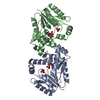
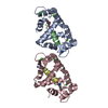
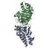
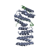
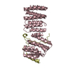
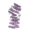
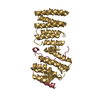
- PDB-2wpv: Crystal structure of S. cerevisiae Get4-Get5 complex -

+
Open data


ID or keywords:

Loading...

-
Basic information

Entry
Database: PDB / ID: 2wpv
TitleCrystal structure of S. cerevisiae Get4-Get5 complex
Components
  • UBIQUITIN-LIKE PROTEIN MDY2
  • UPF0363 PROTEIN YOR164C
KeywordsPROTEIN BINDING / GOLGI-ER TRAFFICKING / TAIL-ANCHORED PROTEIN / GET5 / GET4
Function / homology
Function and homology information


cell morphogenesis involved in conjugation with cellular fusion / TRC complex / tail-anchored membrane protein insertion into ER membrane / protein insertion into ER membrane / post-translational protein targeting to endoplasmic reticulum membrane / vesicle-mediated transport / cytoplasmic stress granule / protein-folding chaperone binding / nucleus / cytoplasm / cytosol
Similarity search - Function
Mdy2, Get4 binding domain / Get5, C-terminal domain / Binding domain to Get4 on Get5, Golgi to ER traffic protein / Ubiquitin-like protein MDY2, C-terminal domain / Golgi to ER traffic protein 4 / Golgi to ER traffic protein 4 / Single helix bin / Tetratricopeptide repeat domain / Single alpha-helices involved in coiled-coils or other helix-helix interfaces / Serine Threonine Protein Phosphatase 5, Tetratricopeptide repeat ...Mdy2, Get4 binding domain / Get5, C-terminal domain / Binding domain to Get4 on Get5, Golgi to ER traffic protein / Ubiquitin-like protein MDY2, C-terminal domain / Golgi to ER traffic protein 4 / Golgi to ER traffic protein 4 / Single helix bin / Tetratricopeptide repeat domain / Single alpha-helices involved in coiled-coils or other helix-helix interfaces / Serine Threonine Protein Phosphatase 5, Tetratricopeptide repeat / Alpha Horseshoe / Ubiquitin family / Ubiquitin homologues / Tetratricopeptide-like helical domain superfamily / Ubiquitin domain profile. / Ubiquitin-like domain / Ubiquitin-like domain superfamily / Up-down Bundle / Mainly Alpha
Similarity search - Domain/homology
: / Golgi to ER traffic protein 4 / Ubiquitin-like protein MDY2
Similarity search - Component
Biological speciesSACCHAROMYCES CEREVISIAE (brewer's yeast)
MethodX-RAY DIFFRACTION / SYNCHROTRON / MAD / Resolution: 1.99 Å
AuthorsChang, Y.-W. / Chuang, Y.-C. / Ho, Y.-C. / Cheng, M.-Y. / Sun, Y.-J. / Hsiao, C.-D. / Wang, C.
CitationJournal: J.Biol.Chem. / Year: 2010
Title: Crystal Structure of Get4-Get5 Complex and its Interactions with Sgt2, Get3, and Ydj1.
Authors: Chang, Y.-W. / Chuang, Y.-C. / Ho, Y.-C. / Cheng, M.-Y. / Sun, Y.-J. / Hsiao, C.-D. / Wang, C.
History
DepositionAug 11, 2009Deposition site: PDBE / Processing site: PDBE
Revision 1.0Jan 26, 2010Provider: repository / Type: Initial release
Revision 1.1Aug 3, 2011Group: Database references / Derived calculations ...Database references / Derived calculations / Other / Refinement description / Structure summary / Version format compliance
Revision 1.2May 8, 2024Group: Data collection / Database references ...Data collection / Database references / Derived calculations / Other
Category: chem_comp_atom / chem_comp_bond ...chem_comp_atom / chem_comp_bond / database_2 / pdbx_database_status / pdbx_struct_conn_angle / struct_conn / struct_site
Item: _database_2.pdbx_DOI / _database_2.pdbx_database_accession ..._database_2.pdbx_DOI / _database_2.pdbx_database_accession / _pdbx_database_status.status_code_sf / _pdbx_struct_conn_angle.ptnr1_auth_comp_id / _pdbx_struct_conn_angle.ptnr1_auth_seq_id / _pdbx_struct_conn_angle.ptnr1_label_atom_id / _pdbx_struct_conn_angle.ptnr1_label_comp_id / _pdbx_struct_conn_angle.ptnr1_label_seq_id / _pdbx_struct_conn_angle.ptnr3_auth_comp_id / _pdbx_struct_conn_angle.ptnr3_auth_seq_id / _pdbx_struct_conn_angle.ptnr3_label_atom_id / _pdbx_struct_conn_angle.ptnr3_label_comp_id / _pdbx_struct_conn_angle.ptnr3_label_seq_id / _struct_conn.pdbx_dist_value / _struct_conn.ptnr1_auth_comp_id / _struct_conn.ptnr1_auth_seq_id / _struct_conn.ptnr1_label_asym_id / _struct_conn.ptnr1_label_atom_id / _struct_conn.ptnr1_label_comp_id / _struct_conn.ptnr1_label_seq_id / _struct_conn.ptnr2_auth_comp_id / _struct_conn.ptnr2_auth_seq_id / _struct_conn.ptnr2_label_asym_id / _struct_conn.ptnr2_label_atom_id / _struct_conn.ptnr2_label_comp_id / _struct_conn.ptnr2_label_seq_id / _struct_site.pdbx_auth_asym_id / _struct_site.pdbx_auth_comp_id / _struct_site.pdbx_auth_seq_id
Remark 650 HELIX DETERMINATION METHOD: AUTHOR PROVIDED.
Remark 700 SHEET DETERMINATION METHOD: AUTHOR PROVIDED.

-
Structure visualization

Structure viewerMolecule:
MolmilJmol/JSmol

Downloads & links

-
Assembly

Deposited unit
A: UPF0363 PROTEIN YOR164C
B: UBIQUITIN-LIKE PROTEIN MDY2
C: UPF0363 PROTEIN YOR164C
D: UBIQUITIN-LIKE PROTEIN MDY2
E: UPF0363 PROTEIN YOR164C
F: UBIQUITIN-LIKE PROTEIN MDY2
G: UPF0363 PROTEIN YOR164C
H: UBIQUITIN-LIKE PROTEIN MDY2
hetero molecules


Theoretical massNumber of molelcules
Total (without water)172,62212
Polymers171,8208
Non-polymers8024
Water21,6541202
1
A: UPF0363 PROTEIN YOR164C
B: UBIQUITIN-LIKE PROTEIN MDY2
hetero molecules


Theoretical massNumber of molelcules
Total (without water)43,1563
Polymers42,9552
Non-polymers2011
Water362
TypeNameSymmetry operationNumber
identity operation1_555x,y,z1
Buried area4840 Å2
ΔGint-61.7 kcal/mol
Surface area15450 Å2
MethodPISA
2
C: UPF0363 PROTEIN YOR164C
D: UBIQUITIN-LIKE PROTEIN MDY2
hetero molecules


Theoretical massNumber of molelcules
Total (without water)43,1563
Polymers42,9552
Non-polymers2011
Water362
TypeNameSymmetry operationNumber
identity operation1_555x,y,z1
Buried area4830 Å2
ΔGint-58.5 kcal/mol
Surface area15310 Å2
MethodPISA
3
E: UPF0363 PROTEIN YOR164C
F: UBIQUITIN-LIKE PROTEIN MDY2
hetero molecules


Theoretical massNumber of molelcules
Total (without water)43,1563
Polymers42,9552
Non-polymers2011
Water362
TypeNameSymmetry operationNumber
identity operation1_555x,y,z1
Buried area4790 Å2
ΔGint-60.6 kcal/mol
Surface area15130 Å2
MethodPISA
4
G: UPF0363 PROTEIN YOR164C
H: UBIQUITIN-LIKE PROTEIN MDY2
hetero molecules


Theoretical massNumber of molelcules
Total (without water)43,1563
Polymers42,9552
Non-polymers2011
Water362
TypeNameSymmetry operationNumber
identity operation1_555x,y,z1
Buried area4810 Å2
ΔGint-61.8 kcal/mol
Surface area15590 Å2
MethodPISA
Unit cell
Length a, b, c (Å)48.275, 118.770, 168.377
Angle α, β, γ (deg.)90.00, 95.17, 90.00
Int Tables number4
Space group name H-MP1211

-
Components

#1: Protein
UPF0363 PROTEIN YOR164C / GET4


Mass: 36320.137 Da / Num. of mol.: 4
Source method: isolated from a genetically manipulated source
Source: (gene. exp.) SACCHAROMYCES CEREVISIAE (brewer's yeast)
Plasmid: PRSFDUET-1 / Production host: ESCHERICHIA COLI (E. coli) / Strain (production host): BL21(DE3) / References: UniProt: Q12125
#2: Protein
UBIQUITIN-LIKE PROTEIN MDY2 / MATING-DEFICIENT PROTEIN 2 / TRANSLATION MACHINERY-ASSOCIATED PROTEIN 24 / GET5


Mass: 6634.776 Da / Num. of mol.: 4 / Fragment: GET4 BINDING DOMAIN, RESIDUES 1-59
Source method: isolated from a genetically manipulated source
Source: (gene. exp.) SACCHAROMYCES CEREVISIAE (brewer's yeast)
Plasmid: PRSFDUET-1 / Production host: ESCHERICHIA COLI (E. coli) / Strain (production host): BL21(DE3) / References: UniProt: Q12285
#3: Chemical
ChemComp-HG / MERCURY (II) ION


Mass: 200.590 Da / Num. of mol.: 4 / Source method: obtained synthetically / Formula: Hg
#4: Water ChemComp-HOH / water


Mass: 18.015 Da / Num. of mol.: 1202 / Source method: isolated from a natural source / Formula: H2O

-
Experimental details

-
Experiment

ExperimentMethod: X-RAY DIFFRACTION / Number of used crystals: 1

-
Sample preparation

CrystalDensity Matthews: 2.81 Å3/Da / Density % sol: 56 % / Description: NONE
Crystal growDetails: 200 MM DI-AMMONIUM HYDROGEN CITRATE AND 20% (V/V) PEG3350

-
Data collection

DiffractionMean temperature: 293 K
Diffraction sourceSource: SYNCHROTRON / Site: NSRRC / Beamline: BL13B1 / Wavelength: 1.008528
DetectorType: ADSC CCD / Detector: CCD / Date: Feb 6, 2009
RadiationProtocol: MAD / Monochromatic (M) / Laue (L): M / Scattering type: x-ray
Radiation wavelengthWavelength: 1.008528 Å / Relative weight: 1
ReflectionResolution: 1.99→30 Å / Num. obs: 239510 / % possible obs: 99.3 % / Observed criterion σ(I): 2 / Redundancy: 3.1 % / Biso Wilson estimate: 9.3 Å2 / Rmerge(I) obs: 0.07 / Net I/σ(I): 15.3
Reflection shellResolution: 1.99→2.06 Å / Redundancy: 3 % / Rmerge(I) obs: 0.33 / Mean I/σ(I) obs: 3.7 / % possible all: 99.1

-
Processing

Software
NameVersionClassification
CNS1.2refinement
HKL-2000data scaling
SOLVE-RESOLVEphasing
RefinementMethod to determine structure: MAD
Starting model: NONE

Resolution: 1.99→25.59 Å / Rfactor Rfree error: 0.002 / Data cutoff high absF: 54151.53 / Data cutoff low absF: 0 / Isotropic thermal model: RESTRAINED / Cross valid method: THROUGHOUT / σ(F): 2
Details: BULK SOLVENT MODEL USED. RESIDUES 1-11 AND 293-312 OF CHAIN A, 1-6 AND 55-59 OF CHAIN B, 1-10 AND 293-312 OF CHAIN C, 1-6 AND 55-59 OF CHAIN D, 1-12 AND 293-312 OF CHAIN E, 1-6 AND 54-59 OF ...Details: BULK SOLVENT MODEL USED. RESIDUES 1-11 AND 293-312 OF CHAIN A, 1-6 AND 55-59 OF CHAIN B, 1-10 AND 293-312 OF CHAIN C, 1-6 AND 55-59 OF CHAIN D, 1-12 AND 293-312 OF CHAIN E, 1-6 AND 54-59 OF CHAIN F, 1-11 AND 293-312 OF CHAIN G, 1-6 AND 55-59 OF CHAIN H ARE MISSING IN THE ELECTRON DENSITY MAP BECAUSE OF DISORDER. SIDECHAIN OF RESIDUES Y135 OF CHAIN A, E11, K15 AND K26 OF CHAIN B, K15, K25, E31, K89, E128, E183, K225 OF CHAIN C, K36 OF CHAIN D, L13, K15, Q18, R19, K25, E31 AND K225 OF CHAIN E, K53 OF CHAIN F, K12, E183 AND K225 OF CHAIN G, K53 OF CHAIN H.
RfactorNum. reflection% reflectionSelection details
Rfree0.206 9814 4.8 %RANDOM
Rwork0.175 ---
obs0.175 203797 80 %-
Solvent computationSolvent model: FLAT MODEL / Bsol: 52.4025 Å2 / ksol: 0.4 e/Å3
Displacement parametersBiso mean: 23.2 Å2
Baniso -1Baniso -2Baniso -3
1--4.34 Å20 Å21.91 Å2
2---2.72 Å20 Å2
3---7.06 Å2
Refine analyze
FreeObs
Luzzati coordinate error0.23 Å0.19 Å
Luzzati d res low-5 Å
Luzzati sigma a0.14 Å0.12 Å
Refinement stepCycle: LAST / Resolution: 1.99→25.59 Å
ProteinNucleic acidLigandSolventTotal
Num. atoms10795 0 4 1202 12001
Refine LS restraints
Refine-IDTypeDev idealDev ideal target
X-RAY DIFFRACTIONc_bond_d0.005
X-RAY DIFFRACTIONc_bond_d_na
X-RAY DIFFRACTIONc_bond_d_prot
X-RAY DIFFRACTIONc_angle_d
X-RAY DIFFRACTIONc_angle_d_na
X-RAY DIFFRACTIONc_angle_d_prot
X-RAY DIFFRACTIONc_angle_deg1
X-RAY DIFFRACTIONc_angle_deg_na
X-RAY DIFFRACTIONc_angle_deg_prot
X-RAY DIFFRACTIONc_dihedral_angle_d18.8
X-RAY DIFFRACTIONc_dihedral_angle_d_na
X-RAY DIFFRACTIONc_dihedral_angle_d_prot
X-RAY DIFFRACTIONc_improper_angle_d0.68
X-RAY DIFFRACTIONc_improper_angle_d_na
X-RAY DIFFRACTIONc_improper_angle_d_prot
X-RAY DIFFRACTIONc_mcbond_it1.221.5
X-RAY DIFFRACTIONc_mcangle_it1.792
X-RAY DIFFRACTIONc_scbond_it2.372
X-RAY DIFFRACTIONc_scangle_it3.572.5
Refine LS restraints NCSNCS model details: NONE
LS refinement shellResolution: 1.99→2.11 Å / Rfactor Rfree error: 0.006 / Total num. of bins used: 6
RfactorNum. reflection% reflection
Rfree0.217 1146 4.7 %
Rwork0.193 23254 -
obs--57.4 %
Xplor file
Refine-IDSerial noParam fileTopol file
X-RAY DIFFRACTION1PROTEIN_REP.PARAMPROTEIN.TOP
X-RAY DIFFRACTION2ION.PARAMION.TOP
X-RAY DIFFRACTION3WATER_REP.PARAMWATER_REP.TOP

+
About Yorodumi

-
News

-
Feb 9, 2022. New format data for meta-information of EMDB entries

New format data for meta-information of EMDB entries

  • Version 3 of the EMDB header file is now the official format.
  • The previous official version 1.9 will be removed from the archive.

Related info.:EMDB header

External links:wwPDB to switch to version 3 of the EMDB data model

-
Aug 12, 2020. Covid-19 info

Covid-19 info

URL: https://pdbjlvh1.pdbj.org/emnavi/covid19.php

New page: Covid-19 featured information page in EM Navigator.

Related info.:Covid-19 info / Mar 5, 2020. Novel coronavirus structure data

+
Mar 5, 2020. Novel coronavirus structure data

Novel coronavirus structure data

Related info.:Yorodumi Speices / Aug 12, 2020. Covid-19 info

External links:COVID-19 featured content - PDBj / Molecule of the Month (242):Coronavirus Proteases

+
Jan 31, 2019. EMDB accession codes are about to change! (news from PDBe EMDB page)

EMDB accession codes are about to change! (news from PDBe EMDB page)

  • The allocation of 4 digits for EMDB accession codes will soon come to an end. Whilst these codes will remain in use, new EMDB accession codes will include an additional digit and will expand incrementally as the available range of codes is exhausted. The current 4-digit format prefixed with “EMD-” (i.e. EMD-XXXX) will advance to a 5-digit format (i.e. EMD-XXXXX), and so on. It is currently estimated that the 4-digit codes will be depleted around Spring 2019, at which point the 5-digit format will come into force.
  • The EM Navigator/Yorodumi systems omit the EMD- prefix.

Related info.:Q: What is EMD? / ID/Accession-code notation in Yorodumi/EM Navigator

External links:EMDB Accession Codes are Changing Soon! / Contact to PDBj

+
Jul 12, 2017. Major update of PDB

Major update of PDB

  • wwPDB released updated PDB data conforming to the new PDBx/mmCIF dictionary.
  • This is a major update changing the version number from 4 to 5, and with Remediation, in which all the entries are updated.
  • In this update, many items about electron microscopy experimental information are reorganized (e.g. em_software).
  • Now, EM Navigator and Yorodumi are based on the updated data.

External links:wwPDB Remediation / Enriched Model Files Conforming to OneDep Data Standards Now Available in the PDB FTP Archive

-
Yorodumi

Thousand views of thousand structures

  • Yorodumi is a browser for structure data from EMDB, PDB, SASBDB, etc.
  • This page is also the successor to EM Navigator detail page, and also detail information page/front-end page for Omokage search.
  • The word "yorodu" (or yorozu) is an old Japanese word meaning "ten thousand". "mi" (miru) is to see.

Related info.:EMDB / PDB / SASBDB / Comparison of 3 databanks / Yorodumi Search / Aug 31, 2016. New EM Navigator & Yorodumi / Yorodumi Papers / Jmol/JSmol / Function and homology information / Changes in new EM Navigator and Yorodumi

Read more