 Movie
Movie Controller

[English] 日本語
 Yorodumi
- PDB-3m62: Crystal structure of Ufd2 in complex with the ubiquitin-like (UBL... -
+ Open data

- Basic information
| Entry | Database: PDB / ID: 3m62 | ||||||
|---|---|---|---|---|---|---|---|
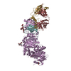
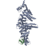
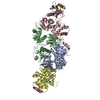
| Title | Crystal structure of Ufd2 in complex with the ubiquitin-like (UBL) domain of Rad23 | ||||||
 Components Components |
| ||||||
 Keywords Keywords | LIGASE/PROTEIN BINDING / Armadillo-like repeats / Ubl conjugation pathway / DNA damage / DNA repair / Nucleus / Phosphoprotein / LIGASE-PROTEIN BINDING complex | ||||||
| Function / homology |  Function and homology information Function and homology informationPNGase complex / nucleotide-excision repair factor 2 complex / nucleotide-excision repair, DNA damage recognition / K48-linked polyubiquitin modification-dependent protein binding / ubiquitin-ubiquitin ligase activity / proteasome binding / Antigen processing: Ubiquitination & Proteasome degradation / cellular response to ethanol / polyubiquitin modification-dependent protein binding / ubiquitin ligase complex ...PNGase complex / nucleotide-excision repair factor 2 complex / nucleotide-excision repair, DNA damage recognition / K48-linked polyubiquitin modification-dependent protein binding / ubiquitin-ubiquitin ligase activity / proteasome binding / Antigen processing: Ubiquitination & Proteasome degradation / cellular response to ethanol / polyubiquitin modification-dependent protein binding / ubiquitin ligase complex / ERAD pathway / protein K48-linked ubiquitination / positive regulation of protein ubiquitination / ubiquitin binding / RING-type E3 ubiquitin transferase / protein polyubiquitination / ubiquitin-dependent protein catabolic process / damaged DNA binding / proteasome-mediated ubiquitin-dependent protein catabolic process / protein-macromolecule adaptor activity / protein ubiquitination / negative regulation of transcription by RNA polymerase II / mitochondrion / nucleoplasm / nucleus / cytoplasm / cytosol Similarity search - Function | ||||||
| Biological species |  | ||||||
| Method |  X-RAY DIFFRACTION / SYNCHROTRON / MOLECULAR REPLACEMENT / molecular replacement / Resolution: 2.4 Å X-RAY DIFFRACTION / SYNCHROTRON / MOLECULAR REPLACEMENT / molecular replacement / Resolution: 2.4 Å | ||||||
 Authors Authors | Haenzelmann, P. / Schindelin, H. | ||||||
 Citation Citation | Journal: J.Biol.Chem. / Year: 2010 Title: The yeast E4 ubiquitin ligase Ufd2 interacts with the ubiquitin-like domains of Rad23 and Dsk2 via a novel and distinct ubiquitin-like binding domain. Authors: Hanzelmann, P. / Stingele, J. / Hofmann, K. / Schindelin, H. / Raasi, S. | ||||||
| History |
|
- Structure visualization
| Structure viewer | Molecule: MolmilJmol/JSmol |
|---|
- Downloads & links
-Download
| PDBx/mmCIF format | 3m62.cif.gz | 226 KB | Display | PDBx/mmCIF format |
|---|---|---|---|---|
| PDB format | pdb3m62.ent.gz | 177 KB | Display | PDB format |
| PDBx/mmJSON format | 3m62.json.gz | Tree view | PDBx/mmJSON format | |
| Others |  Other downloads Other downloads |
-Validation report
| Arichive directory | https://data.pdbj.org/pub/pdb/validation_reports/m6/3m62ftp://data.pdbj.org/pub/pdb/validation_reports/m6/3m62 | HTTPS FTP |
|---|
-Related structure data
| Related structure data |  3m63C  2qizS C: citing same article ( S: Starting model for refinement |
|---|---|
| Similar structure data |


-Links
 PDBj
PDBj


- Assembly
| Deposited unit | 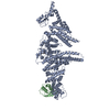
| ||||||||
|---|---|---|---|---|---|---|---|---|---|
| 1 |
| ||||||||
| Unit cell |
|
-Components
| #1: Protein | Mass: 110806.328 Da / Num. of mol.: 1 / Mutation: S102L, D677V Source method: isolated from a genetically manipulated source Source: (gene. exp.) Gene: D1255, UFD2, YDL190C / Production host:  |
|---|---|
| #2: Protein | Mass: 11885.586 Da / Num. of mol.: 1 / Fragment: UNP residues 1-84, Ubiquitin-like domain Source method: isolated from a genetically manipulated source Source: (gene. exp.) Gene: RAD23, SYGP-ORF29, YEL037C / Production host: |
| #3: Chemical | ChemComp-1PE /   Mass: 238.278 Da / Num. of mol.: 1 / Source method: obtained synthetically / Formula: C10H22O6 / Comment: precipitant*YM Mass: 238.278 Da / Num. of mol.: 1 / Source method: obtained synthetically / Formula: C10H22O6 / Comment: precipitant*YM |
| #4: Chemical | ChemComp-K /   Mass: 39.098 Da / Num. of mol.: 1 / Source method: obtained synthetically / Formula: K Mass: 39.098 Da / Num. of mol.: 1 / Source method: obtained synthetically / Formula: K |
| #5: Water | ChemComp-HOH /  Mass: 18.015 Da / Num. of mol.: 298 / Source method: isolated from a natural source / Formula: H2O Mass: 18.015 Da / Num. of mol.: 298 / Source method: isolated from a natural source / Formula: H2O |
-Experimental details
-Experiment
| Experiment | Method: X-RAY DIFFRACTION / Number of used crystals: 1 |
|---|
- Sample preparation
| Crystal | Density Matthews: 3.03 Å3/Da / Density % sol: 59.45 % |
|---|---|
| Crystal grow | Temperature: 298 K / Method: vapor diffusion, hanging drop / pH: 8.3 Details: 16-18% PEG 3500 200 mM Tripotassium citrate, pH 8.3, VAPOR DIFFUSION, HANGING DROP, temperature 298K |
-Data collection
| Diffraction | Mean temperature: 100 K |
|---|---|
| Diffraction source | Source: SYNCHROTRON / Site: BESSY  / Beamline: 14.1 / Wavelength: 0.9 Å / Beamline: 14.1 / Wavelength: 0.9 Å |
| Detector | Type: MARMOSAIC 225 mm CCD / Detector: CCD / Date: Oct 18, 2008 |
| Radiation | Monochromator: Si(111) / Protocol: SINGLE WAVELENGTH / Monochromatic (M) / Laue (L): M / Scattering type: x-ray |
| Radiation wavelength | Wavelength: 0.9 Å / Relative weight: 1 |
| Reflection | Resolution: 2.4→45.22 Å / Num. all: 59314 / Num. obs: 59314 / % possible obs: 100 % / Observed criterion σ(F): 0 / Observed criterion σ(I): 0 / Redundancy: 5.1 % / Rmerge(I) obs: 0.07 / Rsym value: 0.07 / Net I/σ(I): 15.6 |
| Reflection shell | Resolution: 2.4→2.53 Å / Redundancy: 5.2 % / Rmerge(I) obs: 0.492 / Mean I/σ(I) obs: 3.3 / Num. unique all: 59314 / Rsym value: 0.492 / % possible all: 100 |
-Phasing
| Phasing | Method: molecular replacement |
|---|
- Processing
| Software |
| ||||||||||||||||||||||||||||||||||||||||||||||||||||||||||||||||||||||||||||||||||||||||||||||||||||||||||||||||||||||||||||||||||||||||||||||||||||||
|---|---|---|---|---|---|---|---|---|---|---|---|---|---|---|---|---|---|---|---|---|---|---|---|---|---|---|---|---|---|---|---|---|---|---|---|---|---|---|---|---|---|---|---|---|---|---|---|---|---|---|---|---|---|---|---|---|---|---|---|---|---|---|---|---|---|---|---|---|---|---|---|---|---|---|---|---|---|---|---|---|---|---|---|---|---|---|---|---|---|---|---|---|---|---|---|---|---|---|---|---|---|---|---|---|---|---|---|---|---|---|---|---|---|---|---|---|---|---|---|---|---|---|---|---|---|---|---|---|---|---|---|---|---|---|---|---|---|---|---|---|---|---|---|---|---|---|---|---|---|---|---|
| Refinement | Method to determine structure: MOLECULAR REPLACEMENT Starting model: PDB ENTRY 2QIZ Resolution: 2.4→45.22 Å / Cor.coef. Fo:Fc: 0.946 / Cor.coef. Fo:Fc free: 0.911 / Occupancy max: 1 / Occupancy min: 0.5 / SU B: 16.58 / SU ML: 0.177 / TLS residual ADP flag: LIKELY RESIDUAL / Cross valid method: THROUGHOUT / σ(F): 0 / σ(I): 0 / ESU R: 0.307 / ESU R Free: 0.245 / Stereochemistry target values: MAXIMUM LIKELIHOOD Details: HYDROGENS HAVE BEEN ADDED IN THE RIDING POSITIONS U VALUES : RESIDUAL ONLY
| ||||||||||||||||||||||||||||||||||||||||||||||||||||||||||||||||||||||||||||||||||||||||||||||||||||||||||||||||||||||||||||||||||||||||||||||||||||||
| Solvent computation | Ion probe radii: 0.8 Å / Shrinkage radii: 0.8 Å / VDW probe radii: 1.2 Å / Solvent model: MASK | ||||||||||||||||||||||||||||||||||||||||||||||||||||||||||||||||||||||||||||||||||||||||||||||||||||||||||||||||||||||||||||||||||||||||||||||||||||||
| Displacement parameters | Biso max: 87.67 Å2 / Biso mean: 25.733 Å2 / Biso min: 2 Å2
| ||||||||||||||||||||||||||||||||||||||||||||||||||||||||||||||||||||||||||||||||||||||||||||||||||||||||||||||||||||||||||||||||||||||||||||||||||||||
| Refinement step | Cycle: LAST / Resolution: 2.4→45.22 Å
| ||||||||||||||||||||||||||||||||||||||||||||||||||||||||||||||||||||||||||||||||||||||||||||||||||||||||||||||||||||||||||||||||||||||||||||||||||||||
| Refine LS restraints |
| ||||||||||||||||||||||||||||||||||||||||||||||||||||||||||||||||||||||||||||||||||||||||||||||||||||||||||||||||||||||||||||||||||||||||||||||||||||||
| LS refinement shell | Resolution: 2.4→2.462 Å / Total num. of bins used: 20
| ||||||||||||||||||||||||||||||||||||||||||||||||||||||||||||||||||||||||||||||||||||||||||||||||||||||||||||||||||||||||||||||||||||||||||||||||||||||
| Refinement TLS params. | Method: refined / Refine-ID: X-RAY DIFFRACTION
| ||||||||||||||||||||||||||||||||||||||||||||||||||||||||||||||||||||||||||||||||||||||||||||||||||||||||||||||||||||||||||||||||||||||||||||||||||||||
| Refinement TLS group |
|
