 Movie
Movie Controller

[English] 日本語
 Yorodumi
- PDB-5fx8: Complete structure of manganese lipoxygenase of Gaeumannomyces gr... -
+ Open data

- Basic information
| Entry | Database: PDB / ID: 5fx8 | |||||||||
|---|---|---|---|---|---|---|---|---|---|---|
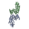
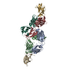
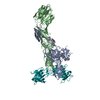
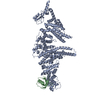
| Title | Complete structure of manganese lipoxygenase of Gaeumannomyces graminis and partial structure of zonadhesin of Komagataella pastoris | |||||||||
 Components Components |
| |||||||||
 Keywords Keywords | OXIDOREDUCTASE / CHIAN A AND B / LINOLEATE 11S-AND 13R- LIPOXYGENASE / FATTTY ACID OXYGENATION / MANGANESE. CHAIN U / FUNGAL ADHESION PROTEIN N-TERMINAL DOMAIN | |||||||||
| Function / homology |  Function and homology information Function and homology informationlinoleate 11-lipoxygenase / linoleate 11-lipoxygenase activity / Oxidoreductases; Acting on single donors with incorporation of molecular oxygen (oxygenases); With incorporation of two atoms of oxygen / lipid oxidation / linoleic acid metabolic process / side of membrane / extracellular region / metal ion binding / plasma membrane Similarity search - Function | |||||||||
| Biological species |  GAEUMANNOMYCES GRAMINIS (fungus)KOMAGATAELLA PHAFFII (fungus) GAEUMANNOMYCES GRAMINIS (fungus)KOMAGATAELLA PHAFFII (fungus) | |||||||||
| Method |  X-RAY DIFFRACTION / SYNCHROTRON / MOLECULAR REPLACEMENT / Resolution: 2.6 Å X-RAY DIFFRACTION / SYNCHROTRON / MOLECULAR REPLACEMENT / Resolution: 2.6 Å | |||||||||
 Authors Authors | Chen, Y. / Wennman, A. / Karkehanadi, S. / Engstrom, A. / Oliw, E.H. | |||||||||
 Citation Citation | Journal: J.Lipid Res. / Year: 2016 Title: Crystal Structure of Linoleate 13R-Manganese Lipoxygenase and an Adhesion Protein Authors: Chen, Y. / Wennman, A. / Karkehabadi, S. / Engstrom, A. / Oliw, E.H. | |||||||||
| History |
|
- Structure visualization
| Structure viewer | Molecule: MolmilJmol/JSmol |
|---|
- Downloads & links
-Download
| PDBx/mmCIF format | 5fx8.cif.gz | 294.3 KB | Display | PDBx/mmCIF format |
|---|---|---|---|---|
| PDB format | pdb5fx8.ent.gz | 236.9 KB | Display | PDB format |
| PDBx/mmJSON format | 5fx8.json.gz | Tree view | PDBx/mmJSON format | |
| Others |  Other downloads Other downloads |
-Validation report
| Arichive directory | https://data.pdbj.org/pub/pdb/validation_reports/fx/5fx8ftp://data.pdbj.org/pub/pdb/validation_reports/fx/5fx8 | HTTPS FTP |
|---|
-Related structure data
| Related structure data |  5fnoS S: Starting model for refinement |
|---|---|
| Similar structure data |

-Links
 PDBj
PDBj
- Assembly
| Deposited unit | 
| ||||||||
|---|---|---|---|---|---|---|---|---|---|
| 1 |
| ||||||||
| Unit cell |
|
-Components
-Protein , 2 types, 3 molecules ABU
| #1: Protein | Mass: 67658.359 Da / Num. of mol.: 2 Source method: isolated from a genetically manipulated source Source: (gene. exp.) GAEUMANNOMYCES GRAMINIS (fungus) / Strain: VAR. AVENAE / Description: CBS 870.03 / Plasmid: PPICZALPHA / Production host: KOMAGATAELLA PASTORIS (fungus) / Strain (production host): X11 / References: UniProt: Q8X151, linoleate 13S-lipoxygenase#2: Protein | | Mass: 34562.527 Da / Num. of mol.: 1 / Fragment: N-TERMINAL DOMAIN / Source method: isolated from a natural source / Details: RESIDUE 20-339 / Source: (natural) KOMAGATAELLA PHAFFII (fungus) / Strain: CBS 7435 / References: UniProt: F2QXM5 |
|---|
-Sugars , 4 types, 10 molecules 
| #3: Polysaccharide | Source method: isolated from a genetically manipulated source #4: Polysaccharide | 2-acetamido-2-deoxy-beta-D-glucopyranose-(1-4)-2-acetamido-2-deoxy-beta-D-glucopyranose-(1-4)-2- ...2-acetamido-2-deoxy-beta-D-glucopyranose-(1-4)-2-acetamido-2-deoxy-beta-D-glucopyranose-(1-4)-2-acetamido-2-deoxy-beta-D-glucopyranose / triacetyl-beta-chitotriose |   Source method: isolated from a genetically manipulated source Details: oligosaccharide / References: triacetyl-beta-chitotriose #5: Polysaccharide | 2-acetamido-2-deoxy-beta-D-glucopyranose-(1-3)-2-acetamido-2-deoxy-beta-D-glucopyranose | Source method: isolated from a genetically manipulated source #7: Sugar | ChemComp-NAG /  Type: D-saccharide, beta linking / Mass: 221.208 Da / Num. of mol.: 5 Type: D-saccharide, beta linking / Mass: 221.208 Da / Num. of mol.: 5Source method: isolated from a genetically manipulated source Formula: C8H15NO6 |
|---|
-Non-polymers , 2 types, 225 molecules 

| #6: Chemical |  Mass: 54.938 Da / Num. of mol.: 2 / Source method: obtained synthetically / Formula: Mn Mass: 54.938 Da / Num. of mol.: 2 / Source method: obtained synthetically / Formula: Mn#8: Water | ChemComp-HOH / | Mass: 18.015 Da / Num. of mol.: 223 / Source method: isolated from a natural source / Formula: H2O |
|---|
-Details
| Has protein modification | Y |
|---|
-Experimental details
-Experiment
| Experiment | Method: X-RAY DIFFRACTION / Number of used crystals: 1 |
|---|
- Sample preparation
| Crystal | Density Matthews: 3 Å3/Da / Density % sol: 50 % / Description: NONE |
|---|---|
| Crystal grow | Details: 0.1M MES-IMIDAZOLE PH6.5, 20%(V/V) GLYCEROL, 10%(W/V)PEG4000, 0.03M MAGNESIUM CHLORIDE, 0.03M CALCIUM CHLORIDE |
-Data collection
| Diffraction | Mean temperature: 100 K |
|---|---|
| Diffraction source | Source: SYNCHROTRON / Site: ESRF  / Beamline: ID29 / Wavelength: 0.976 / Beamline: ID29 / Wavelength: 0.976 |
| Detector | Type: DECTRIS PILATUS 6M / Detector: PIXEL / Date: Jul 29, 2013 |
| Radiation | Protocol: SINGLE WAVELENGTH / Monochromatic (M) / Laue (L): M / Scattering type: x-ray |
| Radiation wavelength | Wavelength: 0.976 Å / Relative weight: 1 |
| Reflection | Resolution: 2.6→49.4 Å / Num. obs: 62790 / % possible obs: 97.8 % / Observed criterion σ(I): 2.4 / Redundancy: 2.9 % / Biso Wilson estimate: 27.74 Å2 / Rmerge(I) obs: 0.1 / Net I/σ(I): 7.6 |
| Reflection shell | Resolution: 2.6→2.8 Å / Redundancy: 3 % / Rmerge(I) obs: 0.75 / Mean I/σ(I) obs: 3 / % possible all: 99 |
- Processing
| Software |
| |||||||||||||||||||||||||||||||||||||||||||||||||||||||||||||||||||||||||||||||||||||||||||||||||||||||||||||||||||||||||||||||||||||||||||||||||||||||||||||||||
|---|---|---|---|---|---|---|---|---|---|---|---|---|---|---|---|---|---|---|---|---|---|---|---|---|---|---|---|---|---|---|---|---|---|---|---|---|---|---|---|---|---|---|---|---|---|---|---|---|---|---|---|---|---|---|---|---|---|---|---|---|---|---|---|---|---|---|---|---|---|---|---|---|---|---|---|---|---|---|---|---|---|---|---|---|---|---|---|---|---|---|---|---|---|---|---|---|---|---|---|---|---|---|---|---|---|---|---|---|---|---|---|---|---|---|---|---|---|---|---|---|---|---|---|---|---|---|---|---|---|---|---|---|---|---|---|---|---|---|---|---|---|---|---|---|---|---|---|---|---|---|---|---|---|---|---|---|---|---|---|---|---|---|
| Refinement | Method to determine structure: MOLECULAR REPLACEMENT Starting model: PDB ENTRY 5FNO Resolution: 2.6→43.163 Å / SU ML: 0.29 / σ(F): 1.34 / Phase error: 23.87 / Stereochemistry target values: ML
| |||||||||||||||||||||||||||||||||||||||||||||||||||||||||||||||||||||||||||||||||||||||||||||||||||||||||||||||||||||||||||||||||||||||||||||||||||||||||||||||||
| Solvent computation | Shrinkage radii: 0.9 Å / VDW probe radii: 1.11 Å / Solvent model: FLAT BULK SOLVENT MODEL | |||||||||||||||||||||||||||||||||||||||||||||||||||||||||||||||||||||||||||||||||||||||||||||||||||||||||||||||||||||||||||||||||||||||||||||||||||||||||||||||||
| Refinement step | Cycle: LAST / Resolution: 2.6→43.163 Å
| |||||||||||||||||||||||||||||||||||||||||||||||||||||||||||||||||||||||||||||||||||||||||||||||||||||||||||||||||||||||||||||||||||||||||||||||||||||||||||||||||
| Refine LS restraints |
| |||||||||||||||||||||||||||||||||||||||||||||||||||||||||||||||||||||||||||||||||||||||||||||||||||||||||||||||||||||||||||||||||||||||||||||||||||||||||||||||||
| LS refinement shell |
|
