 Movie
Movie Controller

[English] 日本語
 Yorodumi
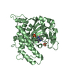
- PDB-6ro8: The crystal structure of Acinetobacter radioresistens CYP116B5 he... -
+ Open data

- Basic information
| Entry | Database: PDB / ID: 6ro8 | ||||||
|---|---|---|---|---|---|---|---|
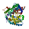
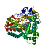
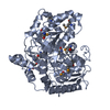
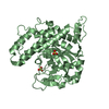
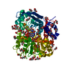
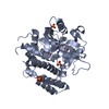
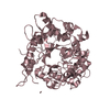
| Title | The crystal structure of Acinetobacter radioresistens CYP116B5 heme domain | ||||||
 Components Components | Cytochrome P450 RhF | ||||||
 Keywords Keywords | OXIDOREDUCTASE / cytochrome P450 / monooxygenases / biocatalysis | ||||||
| Function / homology |  Function and homology information Function and homology informationoxidoreductase activity, acting on paired donors, with incorporation or reduction of molecular oxygen / monooxygenase activity / 2 iron, 2 sulfur cluster binding / iron ion binding / heme binding Similarity search - Function | ||||||
| Biological species |  Acinetobacter radioresistens (bacteria) Acinetobacter radioresistens (bacteria) | ||||||
| Method |  X-RAY DIFFRACTION / SYNCHROTRON / MOLECULAR REPLACEMENT / Resolution: 2.6 Å X-RAY DIFFRACTION / SYNCHROTRON / MOLECULAR REPLACEMENT / Resolution: 2.6 Å | ||||||
 Authors Authors | Ciaramella, A. / Catucci, G. / Gilardi, G. / Di Nardo, G. | ||||||
 Citation Citation | Journal: Int.J.Biol.Macromol. / Year: 2019 Title: Crystal structure of bacterial CYP116B5 heme domain: New insights on class VII P450s structural flexibility and peroxygenase activity. Authors: Ciaramella, A. / Catucci, G. / Gilardi, G. / Di Nardo, G. | ||||||
| History |
|
- Structure visualization
| Structure viewer | Molecule: MolmilJmol/JSmol |
|---|
- Downloads & links
-Download
| PDBx/mmCIF format | 6ro8.cif.gz | 101 KB | Display | PDBx/mmCIF format |
|---|---|---|---|---|
| PDB format | pdb6ro8.ent.gz | 76.3 KB | Display | PDB format |
| PDBx/mmJSON format | 6ro8.json.gz | Tree view | PDBx/mmJSON format | |
| Others |  Other downloads Other downloads |
-Validation report
| Arichive directory | https://data.pdbj.org/pub/pdb/validation_reports/ro/6ro8ftp://data.pdbj.org/pub/pdb/validation_reports/ro/6ro8 | HTTPS FTP |
|---|
-Related structure data
| Related structure data |  6giiS S: Starting model for refinement |
|---|---|
| Similar structure data |

-Links
 PDBj
PDBj


- Assembly
| Deposited unit | 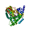
| ||||||||||
|---|---|---|---|---|---|---|---|---|---|---|---|
| 1 |
| ||||||||||
| Unit cell |
|
-Components
| #1: Protein | Mass: 51896.488 Da / Num. of mol.: 1 Source method: isolated from a genetically manipulated source Source: (gene. exp.) Acinetobacter radioresistens (bacteria)Production host: |
|---|---|
| #2: Chemical | ChemComp-HEM /   Mass: 616.487 Da / Num. of mol.: 1 / Source method: obtained synthetically / Formula: C34H32FeN4O4 Mass: 616.487 Da / Num. of mol.: 1 / Source method: obtained synthetically / Formula: C34H32FeN4O4 |
| #3: Chemical | ChemComp-HIS /   Type: L-peptide linking / Mass: 156.162 Da / Num. of mol.: 1 / Source method: obtained synthetically / Formula: C6H10N3O2 Type: L-peptide linking / Mass: 156.162 Da / Num. of mol.: 1 / Source method: obtained synthetically / Formula: C6H10N3O2 |
| #4: Water | ChemComp-HOH /  Mass: 18.015 Da / Num. of mol.: 71 / Source method: isolated from a natural source / Formula: H2O Mass: 18.015 Da / Num. of mol.: 71 / Source method: isolated from a natural source / Formula: H2O |
| Has ligand of interest | N |
-Experimental details
-Experiment
| Experiment | Method: X-RAY DIFFRACTION / Number of used crystals: 1 |
|---|
- Sample preparation
| Crystal | Density Matthews: 5.19 Å3/Da / Density % sol: 76.28 % |
|---|---|
| Crystal grow | Temperature: 277 K / Method: vapor diffusion, sitting drop / pH: 8.5 Details: 100 mM amino acids (0.02 M DL-Arginine hydrochloride, 0.02M DL-Threonine, 0.02M DL-Histidine monohydrochloride monohydrate, 0.02M DL-5-Hydroxylysine hydrochloride, 0.02M trans-4-hydroxy-L- ...Details: 100 mM amino acids (0.02 M DL-Arginine hydrochloride, 0.02M DL-Threonine, 0.02M DL-Histidine monohydrochloride monohydrate, 0.02M DL-5-Hydroxylysine hydrochloride, 0.02M trans-4-hydroxy-L-proline), 0.1 M Gly-Gly, AMPD buffer pH 8.5, 50% v/v of 25% w/v PEG 4000, 40% w/v 1,2,6- Hexanetriol |
-Data collection
| Diffraction | Mean temperature: 100 K / Serial crystal experiment: N |
|---|---|
| Diffraction source | Source: SYNCHROTRON / Site: ESRF  / Beamline: ID23-2 / Wavelength: 1 Å / Beamline: ID23-2 / Wavelength: 1 Å |
| Detector | Type: DECTRIS PILATUS3 X 2M / Detector: PIXEL / Date: Jul 7, 2018 |
| Radiation | Protocol: SINGLE WAVELENGTH / Monochromatic (M) / Laue (L): M / Scattering type: x-ray |
| Radiation wavelength | Wavelength: 1 Å / Relative weight: 1 |
| Reflection | Resolution: 2.6→109.9 Å / Num. obs: 31654 / % possible obs: 100 % / Redundancy: 13.5 % / Biso Wilson estimate: 73.18 Å2 / CC1/2: 1 / Rmerge(I) obs: 0.084 / Net I/av σ(I): 19.9 / Net I/σ(I): 19.9 |
| Reflection shell | Resolution: 2.6→2.72 Å / Redundancy: 12.1 % / Mean I/σ(I) obs: 1.5 / Num. unique obs: 3788 / CC1/2: 0.584 / % possible all: 100 |
- Processing
| Software |
| ||||||||||||||||||||
|---|---|---|---|---|---|---|---|---|---|---|---|---|---|---|---|---|---|---|---|---|---|
| Refinement | Method to determine structure: MOLECULAR REPLACEMENT Starting model: 6GII Resolution: 2.6→91.33 Å / Cross valid method: THROUGHOUT
| ||||||||||||||||||||
| Displacement parameters | Biso max: 202.59 Å2 / Biso mean: 88.6208 Å2 / Biso min: 30 Å2 | ||||||||||||||||||||
| Refinement step | Cycle: LAST / Resolution: 2.6→91.33 Å
|
