 Movie
Movie Controller

[English] 日本語
 Yorodumi
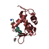
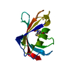
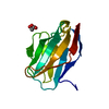
- PDB-6rbb: CRYSTAL STRUCTURE OF the VhH-domain of anti-IL-17A antibody netakimab -
+ Open data

- Basic information
| Entry | Database: PDB / ID: 6rbb | ||||||
|---|---|---|---|---|---|---|---|
| Title | CRYSTAL STRUCTURE OF the VhH-domain of anti-IL-17A antibody netakimab | ||||||
 Components Components | VHH domain of netakimab | ||||||
 Keywords Keywords | IMMUNE SYSTEM / VHH / antibody netakimab / complementarity determining regions / IL-17A / psoriasis | ||||||
| Biological species |  | ||||||
| Method |  X-RAY DIFFRACTION / SYNCHROTRON / MOLECULAR REPLACEMENT / Resolution: 2.45 Å X-RAY DIFFRACTION / SYNCHROTRON / MOLECULAR REPLACEMENT / Resolution: 2.45 Å | ||||||
 Authors Authors | Kostareva, O.S. / Kolyadenko, I.A. / Ulitin, A.B. / Ekimova, V.M. / Evdokimov, S.R. / Garber, M.B. / Tishchenko, T.V. / Gabdulkhakov, A.G. | ||||||
| Funding support |  Russian Federation, 1items Russian Federation, 1items
| ||||||
 Citation Citation | Journal: To Be Published Title: CRYSTAL STRUCTURE OF the VhH-domain of anti-IL-17A antibody netakimab Authors: Kostareva, O.S. / Kolyadenko, I.A. / Ulitin, A.B. / Ekimova, V.M. / Evdokimov, S.R. / Garber, M.B. / Tishchenko, T.V. / Gabdulkhakov, A.G. | ||||||
| History |
|
- Structure visualization
| Structure viewer | Molecule:  MolmilJmol/JSmol MolmilJmol/JSmol |
|---|
- Downloads & links
-Download
| PDBx/mmCIF format | 6rbb.cif.gz | 102 KB | Display | PDBx/mmCIF format |
|---|---|---|---|---|
| PDB format | pdb6rbb.ent.gz | 78.9 KB | Display | PDB format |
| PDBx/mmJSON format | 6rbb.json.gz | Tree view | PDBx/mmJSON format | |
| Others |  Other downloads Other downloads |
-Validation report
| Arichive directory | https://data.pdbj.org/pub/pdb/validation_reports/rb/6rbbftp://data.pdbj.org/pub/pdb/validation_reports/rb/6rbb | HTTPS FTP |
|---|
-Related structure data
| Related structure data |  6qkdS S: Starting model for refinement |
|---|---|
| Similar structure data |


-Links
 PDBj
PDBj

- Assembly
| Deposited unit | 
| ||||||||
|---|---|---|---|---|---|---|---|---|---|
| 1 | 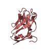
| ||||||||
| 2 | 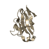
| ||||||||
| 3 | 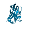
| ||||||||
| 4 | 
| ||||||||
| Unit cell |
|
-Components
| #1: Antibody | Mass: 13241.626 Da / Num. of mol.: 4 Source method: isolated from a genetically manipulated source Source: (gene. exp.)  Cricetulus griseus (Chinese hamster) Cricetulus griseus (Chinese hamster)#2: Water | ChemComp-HOH / |  Mass: 18.015 Da / Num. of mol.: 26 / Source method: isolated from a natural source / Formula: H2O Mass: 18.015 Da / Num. of mol.: 26 / Source method: isolated from a natural source / Formula: H2OHas protein modification | Y | |
|---|
-Experimental details
-Experiment
| Experiment | Method: X-RAY DIFFRACTION / Number of used crystals: 1 |
|---|
- Sample preparation
| Crystal | Density Matthews: 2.43 Å3/Da / Density % sol: 49.37 % |
|---|---|
| Crystal grow | Temperature: 296 K / Method: vapor diffusion, hanging drop / pH: 5 Details: 50mMNa AcpH 5.5, 150mMNaCl with 0.1M Citrate pH 5, 20% (w/v) PEG 6000 |
-Data collection
| Diffraction | Mean temperature: 100 K / Serial crystal experiment: N |
|---|---|
| Diffraction source | Source: SYNCHROTRON / Site: ESRF  / Beamline: ID23-1 / Wavelength: 1.0332 Å / Beamline: ID23-1 / Wavelength: 1.0332 Å |
| Detector | Type: DECTRIS PILATUS3 S 6M / Detector: PIXEL / Date: Oct 27, 2018 |
| Radiation | Protocol: SINGLE WAVELENGTH / Monochromatic (M) / Laue (L): M / Scattering type: x-ray |
| Radiation wavelength | Wavelength: 1.0332 Å / Relative weight: 1 |
| Reflection | Resolution: 2.38→50 Å / Num. obs: 21024 / % possible obs: 98.3 % / Redundancy: 5.1 % / CC1/2: 0.996 / Rrim(I) all: 0.154 / Net I/σ(I): 9.1 |
| Reflection shell | Resolution: 2.38→2.52 Å / Mean I/σ(I) obs: 1.4 / Num. unique obs: 3076 / % possible all: 90.7 |
- Processing
| Software |
| ||||||||||||||||||||||||||||||||||||||||||||||||||||||||||||||||||||||||||||||||||||||||||||||||||
|---|---|---|---|---|---|---|---|---|---|---|---|---|---|---|---|---|---|---|---|---|---|---|---|---|---|---|---|---|---|---|---|---|---|---|---|---|---|---|---|---|---|---|---|---|---|---|---|---|---|---|---|---|---|---|---|---|---|---|---|---|---|---|---|---|---|---|---|---|---|---|---|---|---|---|---|---|---|---|---|---|---|---|---|---|---|---|---|---|---|---|---|---|---|---|---|---|---|---|---|
| Refinement | Method to determine structure: MOLECULAR REPLACEMENT Starting model: 6QKD Resolution: 2.45→41.846 Å / SU ML: 0.34 / Cross valid method: FREE R-VALUE / σ(F): 0.02 / Phase error: 32.24
| ||||||||||||||||||||||||||||||||||||||||||||||||||||||||||||||||||||||||||||||||||||||||||||||||||
| Solvent computation | Shrinkage radii: 0.9 Å / VDW probe radii: 1.11 Å | ||||||||||||||||||||||||||||||||||||||||||||||||||||||||||||||||||||||||||||||||||||||||||||||||||
| Refinement step | Cycle: LAST / Resolution: 2.45→41.846 Å
| ||||||||||||||||||||||||||||||||||||||||||||||||||||||||||||||||||||||||||||||||||||||||||||||||||
| Refine LS restraints |
| ||||||||||||||||||||||||||||||||||||||||||||||||||||||||||||||||||||||||||||||||||||||||||||||||||
| LS refinement shell |
|
